Accéder au contenu
Merck
Toutes les photos(1)

Key Documents

358M-1

Sigma-Aldrich

MUM1 (MRQ-8) Mouse Monoclonal Antibody

Se connecterpour consulter vos tarifs contractuels et ceux de votre entreprise/organisme


About This Item

Code UNSPSC :
12352203
Nomenclature NACRES :
NA.41

Source biologique

mouse

Niveau de qualité

100
500

Conjugué

unconjugated

Forme d'anticorps

culture supernatant

Type de produit anticorps

primary antibodies

Clone

MRQ-8, monoclonal

Description

For In Vitro Diagnostic Use in Select Regions (See Chart)

Forme

buffered aqueous solution

Espèces réactives

human

Conditionnement

vial of 0.1 mL concentrate (358M-14)
vial of 0.5 mL concentrate (358M-15)
bottle of 1.0 mL predilute (358M-17)
vial of 1.0 mL concentrate (358M-16)
bottle of 7.0 mL predilute (358M-18)

Fabricant/nom de marque

Cell Marque

Technique(s)

immunohistochemistry (formalin-fixed, paraffin-embedded sections): 1:100-1:500

Isotype

IgG1κ

Contrôle

B-cell chronic lymphocytic leukemia (B-CLL), plasma cell myeloma, tonsil

Conditions d'expédition

wet ice

Température de stockage

2-8°C

Visualisation

cytoplasmic, nuclear

Informations sur le gène

human ... PWWP3A(84939)

Description générale

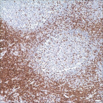
MUM1 (multiple myeloma oncogene-1)/IRF4 (interferon regulatory factor 4) is a 50 kDa protein encoded by MUM1 gene, and a member of the interferon regulatory factor family of transcription factors. MUM1/IRF4 is expressed in the nuclei of plasma cells and a small percentage of germinal center (GC) B-cells located in the “light zone”. This antibody stains MUM1 protein, which is expressed in a subset of B-cells in the light zone of the germinal center, plasma cells, activated T-cells, and a wide spectrum of related hematolymphoid neoplasms derived from these cells. Therefore, this antibody is useful for the subclassiffication of lymphoid malignancies and an excellent marker for Reed-Sternberg cells of classic Hodgkin disease in combination with anti-CD30.

Qualité


IVD

IVD

IVD

RUO

Liaison

MUM1 Positive Control Slides, Product No. 358S, are available for immunohistochemistry (formalin-fixed, paraffin-embedded sections).

Forme physique

Solution in Tris Buffer, pH 7.3-7.7, with 1% BSA and <0.1% Sodium Azide

Notes préparatoires

Download the IFU specific to your product lot and formatNote: This requires a keycode which can be found on your packaging or product label.

Autres remarques

For Technical Service please contact: 800-665-7284 or email: service@cellmarque.com

Informations légales

Cell Marque is a trademark of Merck KGaA, Darmstadt, Germany

Vous ne trouvez pas le bon produit ?  

Essayez notre Outil de sélection de produits.


Certificats d'analyse (COA)

Recherchez un Certificats d'analyse (COA) en saisissant le numéro de lot du produit. Les numéros de lot figurent sur l'étiquette du produit après les mots "Lot" ou "Batch".

Déjà en possession de ce produit ?

Retrouvez la documentation relative aux produits que vous avez récemment achetés dans la Bibliothèque de documents.

Consulter la Bibliothèque de documents

G Gaidano et al.
Leukemia, 14(4), 563-566 (2000-04-14)
In recent times, the field of B cell lymphoma histogenesis has progressed rapidly due to the increasing availability of histogenetic markers. Genotypic markers of B cell histogenesis are represented by mutations of IgV and BCL-6 genes, which are somatically acquired
Y Natkunam et al.
Modern pathology : an official journal of the United States and Canadian Academy of Pathology, Inc, 14(7), 686-694 (2001-07-17)
The gene encoding MUM1 was characterized as a possible translocation partner in chromosomal abnormalities involving a significant number of multiple myelomas. The overexpression of the MUM1 protein as a result of translocation t(6;14) (p25;q32) identified MUM1 as a putative regulatory
B Falini et al.
Blood, 95(6), 2084-2092 (2000-03-09)
A new monoclonal antibody (MUM1p) was used to study the cell/tissue expression of human MUM1/IRF4 protein, the product of the homologous gene involved in the myeloma-associated t(6;14) (p25;q32). MUM1 was expressed in the nuclei and cytoplasm of plasma cells and
Antonino Carbone et al.
British journal of haematology, 117(2), 366-372 (2002-04-26)
Biological and clinical studies have shown that Hodgkin's disease (HD) can be divided into two major categories, termed nodular lymphocyte predominance HD (NLP HD) and classic HD (CHD). Within CHD four subtypes have been distinguished: nodular sclerosis, mixed cellularity, lymphocyte
Mathewos Tessema et al.
PloS one, 7(4), e34850-e34850 (2012-04-13)
Aberrant cytosine methylation affects regulation of hundreds of genes during cancer development. In this study, a novel aberrantly hypermethylated CpG island in cancer was discovered within the TOX2 promoter. TOX2 was unmethylated in normal cells but 28% lung (n =

Notre équipe de scientifiques dispose d'une expérience dans tous les secteurs de la recherche, notamment en sciences de la vie, science des matériaux, synthèse chimique, chromatographie, analyse et dans de nombreux autres domaines..

Contacter notre Service technique