Accéder au contenu
Merck
Toutes les photos(2)

Key Documents

CBL218-I

Sigma-Aldrich

Anti-Keratin K3/K76 Antibody, clone AE5

clone AE5, from mouse

Synonyme(s) :

65 kDa cytokeratin, Cytokeratin-3, CK-3, Keratin-3, K3, Type-II keratin Kb3

Se connecterpour consulter vos tarifs contractuels et ceux de votre entreprise/organisme


About This Item

Code UNSPSC :
12352203
eCl@ss :
32160702
Nomenclature NACRES :
NA.41

Source biologique

mouse

Niveau de qualité

Forme d'anticorps

purified immunoglobulin

Type de produit anticorps

primary antibodies

Clone

AE5, monoclonal

Espèces réactives

rabbit, bovine

Technique(s)

immunofluorescence: suitable
immunohistochemistry: suitable
western blot: suitable

Isotype

IgG1κ

Numéro d'accès NCBI

Numéro d'accès UniProt

Conditions d'expédition

wet ice

Modification post-traductionnelle de la cible

unmodified

Informations sur le gène

human ... KRT3(3850)

Description générale

Cytokeratin 3, also known as 65kDa Cytokeratin or Keratin K3 is a specific keratin found primarily in the corneal epithelium and minorly in other epithelium like that of the esophagus. The corneal epithelium tissue forms the outermost layer of the cornea, which is the clear front covering of the eye. Keratins are divided into two primary types based upon their isoelectric mobility and size, acidic, or type 1:40-55kDa, and basic to neutral or type 2, 56-70kDa. Cytokeratin 3 is a type II cytokeratin. Type II cytokeratins form pairs with other cytokeratins. Cytokeratin 3 is expressed in the corneal epithelium with Cytokeratin 12. Together Cytokeratin 3 and Cytokeratin 12 form molecules known as intermediate filaments. These filaments assemble into strong networks that provide strength and resilience to the corneal epithelium. ′

Immunogène

Whole cells corresponding to Rabbit Keratin, type II cytosleletal 3.

Application

Analyse par immunohistochimie :
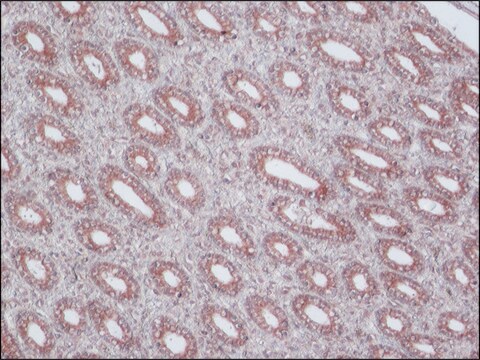
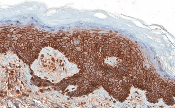
A representative lot was used by an independent laboratory in Keratin 3 expressed epithelial cells on the surface of submerged and AL explants, indicating that they were derived from limbal progenitor cells via migration.
A representative lot was used by an independent laboratory in Rabbit epithelial Keratin lysate. (Schermer, A, et al. (1986) J. Cell Biol., 103:
A representative lot was used by an independent laboratory in rabbit corneal tissue lysate. (Cooper, D., et al. (1986) J. Biol. Chem., 261:
A representative lot was used by an independent laboratory in human limbal stem cells cultured in medium supplemented with human EGF. (Sharifi, A.M., et al. (2010) Biocell.
A representative lot was used by an independent laboratory in rabbit corneal and limbal tissues, limbal explant, and epithelial outgrowth on human AM.
A representative lot was used by an independent laboratory inRabbit corneal epithelial Keratin cells. (Schermer, A, et al. (1986) J. Cell Biol., 103: 49-62).
Domaine de recherche
Structure cellulaire
Sous-domaine de recherche
Cytokératines
This Anti-Keratin K3/K76 Antibody, clone AE5 is validated for use in Western Blotting and Immunohistochemistry and Immunofluorescence for the detection of Keratin K3/K76.

Qualité


µµ

Description de la cible

Env. 60 kDa (taille observée)

Liaison

Remplace le(s) produit(s) suivant(s) : CBL218

Forme physique

Format : Produit purifié
IgG1κ monoclonale de souris purifiée, dans un tampon à base de Tris-glycine 0,1 M (pH 7,4) et de NaCl 150 mM contenant 0,05 % d'azoture de sodium.
Produit purifié sur protéine G

Stockage et stabilité

Stable entre 2 et 8 °C pendant 1 an à compter de la date de réception.

Autres remarques

Concentration : Voir la fiche technique du lot concerné.

Clause de non-responsabilité

Sauf indication contraire dans notre catalogue ou toute autre documentation associée au(x) produit(s), nos produits sont uniquement destinés à la recherche et ne sauraient être utilisés à d'autres fins, ce qui inclut, sans s'y limiter, les utilisations commerciales non autorisées, les utilisations diagnostiques in vitro, les utilisations thérapeutiques ex vivo ou in vivo, ou tout type de consommation ou d'application chez l'être humain ou chez l'animal.

Vous ne trouvez pas le bon produit ?  

Essayez notre Outil de sélection de produits.

Code de la classe de stockage

12 - Non Combustible Liquids

Classe de danger pour l'eau (WGK)

WGK 1

Point d'éclair (°F)

Not applicable

Point d'éclair (°C)

Not applicable


Certificats d'analyse (COA)

Recherchez un Certificats d'analyse (COA) en saisissant le numéro de lot du produit. Les numéros de lot figurent sur l'étiquette du produit après les mots "Lot" ou "Batch".

Déjà en possession de ce produit ?

Retrouvez la documentation relative aux produits que vous avez récemment achetés dans la Bibliothèque de documents.

Consulter la Bibliothèque de documents

Differentiation-related expression of a major 64K corneal keratin in vivo and in culture suggests limbal location of corneal epithelial stem cells.
Schermer, A; Galvin, S; Sun, TT
The Journal of cell biology null
Intrastromal invasion by limbal epithelial cells is mediated by epithelial-mesenchymal transition activated by air exposure.
Kawakita, T; Espana, EM; He, H; Li, W; Liu, CY; Tseng, SC
The American Journal of Pathology null
Monoclonal antibody analysis of bovine epithelial keratins. Specific pairs as defined by coexpression.
Cooper, D and Sun, T T
The Journal of Biological Chemistry, 261, 4646-4654 (1986)
Propagation and phenotypic preservation of rabbit limbal epithelial cells on amniotic membrane.
Wang, DY; Hsueh, YJ; Yang, VC; Chen, JK
Investigative Ophthalmology & Visual Science null

Notre équipe de scientifiques dispose d'une expérience dans tous les secteurs de la recherche, notamment en sciences de la vie, science des matériaux, synthèse chimique, chromatographie, analyse et dans de nombreux autres domaines..

Contacter notre Service technique