Accéder au contenu
Merck
Toutes les photos(2)

Principaux documents

ABF210

Sigma-Aldrich

Anti-MDA5 Antibody

from rabbit, purified by affinity chromatography

Synonyme(s) :

MDA5 Antibody, Melanoma differentiation-associated protein 5, Interferon-induced with helicase C domain protein 1, IFIH1

Se connecterpour consulter vos tarifs contractuels et ceux de votre entreprise/organisme


About This Item

Code UNSPSC :
12352203
eCl@ss :
32160702
Nomenclature NACRES :
NA.41

Source biologique

rabbit

Niveau de qualité

Forme d'anticorps

affinity isolated antibody

Type de produit anticorps

primary antibodies

Clone

polyclonal

Produit purifié par

affinity chromatography

Espèces réactives

human

Technique(s)

immunofluorescence: suitable
immunohistochemistry: suitable (paraffin)
western blot: suitable

Numéro d'accès UniProt

Conditions d'expédition

wet ice

Modification post-traductionnelle de la cible

unmodified

Informations sur le gène

human ... IFIH1(64135)

Description générale

Melanoma differentiation-associated protein 5 (MDA5), also known as Interferon-induced helicase C domain-containing protein 1 (IFIH1) is a member of the helicase family (RLR subfamily). MDA5 plays an important role in recognizing various viral components and triggers antiviral responses. When stimulated by dsRNA, MDA5 recruits adaptor protein VISA causing the activation of IRF-3 and NF-κB. MDA5 is widely expressed at low levels with barely detectable levels found in , testis and lung. Mutations in MDA5 have been associated with diabetes mellitus insulin-dependent type 19.

Spécificité

This antibody recognizes the C-terminus of MDA5.

Immunogène

Epitope: C-terminus
KLH-conjugated linear peptide corresponding to the C-terminus of human MDA5.

Application

Detect Melanoma differentiation-associated protein 5 using this rabbit polyclonal antibody, Anti-MDA5 Antibody validated for use in western blotting, IHC (Paraffin) & Immunofluorescence.
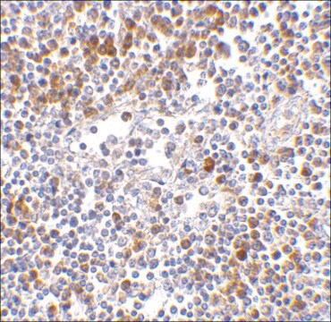
Immunohistochemistry Analysis: 5 µg/mL from a representative lot detected MDA5 in human lymph node tissue lysate.

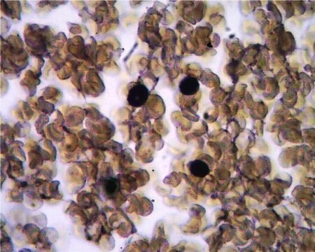
Immunofluorescence Analysis: 20 μg/mL from a representative lot detected MDA5 in human lymph node cells.
Research Category
Inflammation & Immunology
Research Sub Category
Immunoglobulins & Immunology

Qualité

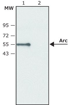
Evaluated by Western Blotting in Daudi cell lysate.

Western Blotting Analysis: 2 µg/mL of this antibody detected MDA5 in 15 µg of Daudi cell lysate.

Description de la cible

~117 kDa observed. Uncharacterized bands may appear in some lysates.

Liaison

Replaces: MABF198

Forme physique

Antigen Affinity Purified
Purified rabbit polyclonal in buffer containing PBS with up to 0.1% sodium azide.

Stockage et stabilité

Stable for 1 year at 2-8°C from date of receipt.

Remarque sur l'analyse

Control
Daudi cell Lysate

Autres remarques

Concentration: Please refer to the Certificate of Analysis for the lot-specific concentration.

Clause de non-responsabilité

Unless otherwise stated in our catalog or other company documentation accompanying the product(s), our products are intended for research use only and are not to be used for any other purpose, which includes but is not limited to, unauthorized commercial uses, in vitro diagnostic uses, ex vivo or in vivo therapeutic uses or any type of consumption or application to humans or animals.

Vous ne trouvez pas le bon produit ?  

Essayez notre Outil de sélection de produits.

Code de la classe de stockage

12 - Non Combustible Liquids

Classe de danger pour l'eau (WGK)

WGK 2

Point d'éclair (°F)

Not applicable

Point d'éclair (°C)

Not applicable


Certificats d'analyse (COA)

Recherchez un Certificats d'analyse (COA) en saisissant le numéro de lot du produit. Les numéros de lot figurent sur l'étiquette du produit après les mots "Lot" ou "Batch".

Déjà en possession de ce produit ?

Retrouvez la documentation relative aux produits que vous avez récemment achetés dans la Bibliothèque de documents.

Consulter la Bibliothèque de documents

Ling Xu et al.
Proceedings of the National Academy of Sciences of the United States of America, 113(39), 10950-10955 (2016-09-14)
The function of the RIG-I-like receptors (RLRs; including RIG-I, MDA5, and LGP2) as key cytoplasmic sensors of viral pathogen-associated molecular patterns (PAMPs) has been subjected to numerous pathogenic challenges and has undergone a dynamic evolution. We found evolutionary evidence that

Notre équipe de scientifiques dispose d'une expérience dans tous les secteurs de la recherche, notamment en sciences de la vie, science des matériaux, synthèse chimique, chromatographie, analyse et dans de nombreux autres domaines..

Contacter notre Service technique