Skip to Content
Merck
All Photos(2)

Key Documents

SAB1400139

Sigma-Aldrich

Anti-IL6 antibody produced in mouse

IgG fraction of antiserum, buffered aqueous solution

Synonym(s):

Anti-BSF2, Anti-HGF, Anti-HSF, Anti-IFNB2, Anti-IL6

Sign Into View Organizational & Contract Pricing


About This Item

MDL number:
UNSPSC Code:
12352203
NACRES:
NA.41

biological source

mouse

conjugate

unconjugated

antibody form

IgG fraction of antiserum

antibody product type

primary antibodies

clone

polyclonal

form

buffered aqueous solution

species reactivity

human

technique(s)

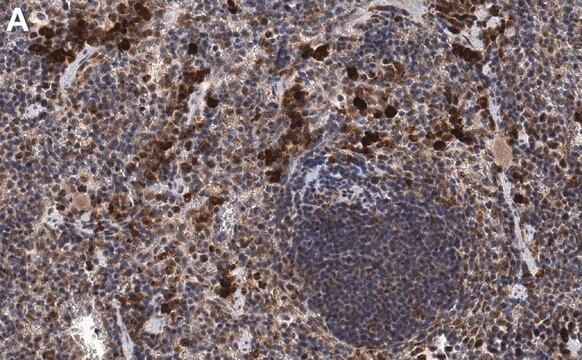
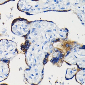
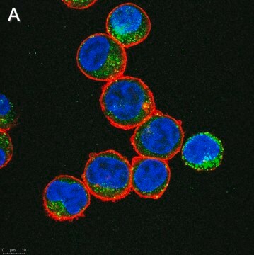
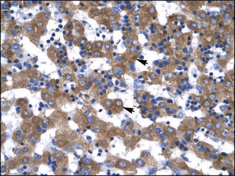
immunofluorescence: suitable
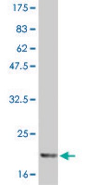
western blot: 1 μg/mL

UniProt accession no.

shipped in

dry ice

storage temp.

−20°C

target post-translational modification

unmodified

Gene Information

human ... IL6(3569)

General description

Human IL-6 (Interleukin-6) gene maps to chromosome 7p15. It is found to be mainly expressed in lymphoid and non-lymphoid cells, such as T-cells, B-cells, monocytes, fibroblasts, keratinocytes, endothelial cells and mesangium cells. It encodes a 184 amino acid protein that contains two potential N-glycosylation sites and four cysteine residues.

This gene encodes a cytokine that functions in inflammation and the maturation of B cells. The protein is primarily produced at sites of acute and chronic inflammation, where it is secreted into the serum and induces a transcriptional inflammatory response through interleukin 6 receptor, alpha. The functioning of this gene is implicated in a wide variety of inflammation-associated disease states, including suspectibility to diabetes mellitus and systemic juvenile rheumatoid arthritis. (provided by RefSeq)

Immunogen

IL6 (AAH15511, 1 a.a. ~ 212 a.a) full-length human protein.

Sequence
MNSFSTSAFGPVAFSLGLLLVLPAAFPAPVPPGEDSKDVAAPHRQPLTSSERIDKQIRYILDGISALRKETCNKSNMCESSKEALAENNLNLPKMAEKDGCFQSGFNEETCLVKIITGLLEFEVYLEYLQNRFESSEEQARAVQMSTKVLIQFLQKKAKNLDAITTPDPTTNASLLTKLQAQNQWLQDMTTHLILRSFKEFLQSSLRALRQM

Biochem/physiol Actions

IL-6 (Interleukin-6) is a proinflammatory cytokine that plays an important role in the maturation of B cells into antibody producing cells. It is also expressed by resting T-cells and induces IL-2 receptor and IL-2 production n mitogen-stimulated T cells and thymocytes. IL-6 functions in the activation and proliferation of T-cells. It participates in hematopoiesis by activating hematopoietic stem cells at the G0 stage to enter into the G1 phase. Increased levels of IL-6 and C-reactive protein have been observed in type-2 diabetes mellitus.

Physical form

Solution in phosphate buffered saline, pH 7.4

Disclaimer

Unless otherwise stated in our catalog or other company documentation accompanying the product(s), our products are intended for research use only and are not to be used for any other purpose, which includes but is not limited to, unauthorized commercial uses, in vitro diagnostic uses, ex vivo or in vivo therapeutic uses or any type of consumption or application to humans or animals.

Not finding the right product?  

Try our Product Selector Tool.

Storage Class Code

10 - Combustible liquids

Flash Point(F)

Not applicable

Flash Point(C)

Not applicable


Certificates of Analysis (COA)

Search for Certificates of Analysis (COA) by entering the products Lot/Batch Number. Lot and Batch Numbers can be found on a product’s label following the words ‘Lot’ or ‘Batch’.

Already Own This Product?

Find documentation for the products that you have recently purchased in the Document Library.

Visit the Document Library

The biology of interleukin-6.
Hirano T.
Interleukins : molecular biology and immunology, 51, 153-180 (1992)
The biology of interleukin-6.
Hirano T.
INTERNATIONAL CONFERENCE OF COMPUTATIONAL METHODS IN SCIENCES AND ENGINEERING 2009:(ICCMSE 2009)., 51, 153-180 (1992)
A D Pradhan et al.
JAMA, 286(3), 327-334 (2001-07-24)
Inflammation is hypothesized to play a role in development of type 2 diabetes mellitus (DM); however, clinical data addressing this issue are limited. To determine whether elevated levels of the inflammatory markers interleukin 6 (IL-6) and C-reactive protein (CRP) are
Lorena Oróstica et al.
Reproductive sciences (Thousand Oaks, Calif.), 27(1), 290-300 (2020-02-13)
A pro-inflammatory environment is characteristic of obesity and polycystic ovary syndrome (PCOS). This environment through cytokines secretion negatively affects insulin action. Endometria from women with both conditions (obesity and PCOS) present high TNF-α level and altered insulin signaling. In addition
Hagen Maxeiner et al.
Journal of cellular physiology, 229(11), 1681-1689 (2014-03-14)
Cardiosphere-derived cells (CDCs) were cultured from human, murine, and rat hearts. Diluted supernatant (conditioned-medium) of the cultures improved the contractile behavior of isolated rat cardiomyocytes (CMCs). This effect is mediated by the paracrine release of cytokines. The present study tested

Our team of scientists has experience in all areas of research including Life Science, Material Science, Chemical Synthesis, Chromatography, Analytical and many others.

Contact Technical Service