Skip to Content
Merck
All Photos(3)

Key Documents

PKH26GL

Sigma-Aldrich

PKH26 Red Fluorescent Cell Linker Kit for General Cell Membrane Labeling

Distributed for Phanos Technologies

Synonym(s):

Red PKH membrane dye

Sign Into View Organizational & Contract Pricing


About This Item

UNSPSC Code:
12352207
NACRES:
NA.32

packaging

pkg of 1 kit

Quality Level

manufacturer/tradename

Distributed for Phanos Technologies

storage condition

protect from light

fluorescence

λex 551 nm; λem 567 nm (PKH26 dye)

detection method

fluorometric

shipped in

ambient

storage temp.

room temp

General description

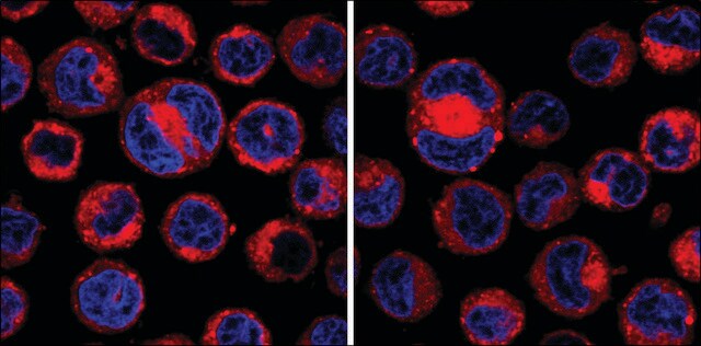
PKH26 is a red‐fluorescent dye. This lipophilic membrane dye is widely used to determine cell proliferation in heterogeneous cells by flow cytometry.

Application

PKH26 Red Fluorescent Cell Linker Kit is for general cell membrane labeling. It has been characterized in a number of model systems and has been found to be useful for in vitro cell labeling, in vitro proliferation studies and long term, in vivo cell tracking. The half-life for elution of PKH26 from labeled rabbit red blood cells is greater than 100 days. This enhanced stability is favorable for long term in vivo studies.
This kit is for general cell membrane labeling. It has been characterized in a number of model systems and has been found to be useful for in vitro cell labeling, in vitro proliferation studies and long term, in vivo cell tracking. The half-life for elution of PKH26 from labeled rabbit red blood cells is greater than 100 days. This enhanced stability is favorable for long term in vivo studies.

Linkage

For additional technical details on PKH and CellVue® Fluorescent Cell Linker Dyes including an extensive bibliography, please visit here.

Legal Information

CellVue is a registered trademark of Phanos Technologies

Kit Components Only

Product No.
Description

  • Diluent C 6 x 10

  • PKH26 Cell Linker in ethanol .5 mL

related product

Pictograms

FlameExclamation mark

Signal Word

Danger

Hazard Statements

Hazard Classifications

Eye Irrit. 2 - Flam. Liq. 2

Storage Class Code

3 - Flammable liquids

WGK

WGK 1

Flash Point(F)

57.2 °F - closed cup

Flash Point(C)

14.0 °C - closed cup


Choose from one of the most recent versions:

Certificates of Analysis (COA)

Lot/Batch Number

Don't see the Right Version?

If you require a particular version, you can look up a specific certificate by the Lot or Batch number.

Already Own This Product?

Find documentation for the products that you have recently purchased in the Document Library.

Visit the Document Library

EphA3 as a target for antibody immunotherapy in acute lymphoblastic leukemia
Charmsaz S, et al.
Leukemia, 31(8), 1779-1779 (2017)
Rafi Chapanian et al.
Biomaterials, 33(10), 3047-3057 (2012-01-21)
The in vivo circulation of hyperbranched polyglycerol (HPG) grafted red blood cells (RBCs) was investigated in mice. The number of HPG molecules grafted per RBC was measured using tritium labeled HPGs ((3)H-HPG) of different molecular weights; the values ranged from
Anne Julie Frenette et al.
Journal of neurotrauma, 29(1), 1-18 (2011-08-19)
In the intensive care unit, dopamine agonists (DA) have been used in traumatic brain injury (TBI) patients to augment or accelerate cognitive recovery and rehabilitation. However, the efficacy and safety of DA in this population is not well established. We
Ofra Ben Menachem-Zidon et al.
PloS one, 14(6), e0218081-e0218081 (2019-06-14)
The beneficial effect of mesenchymal stem cells (MSCs) on wound healing is mostly attributed to a trophic effect that promotes angiogenesis. Whether MSCs can contribute to the formation of new blood vessels by direct differentiation is still controversial. Pelvic floor
Tracking antigen-driven responses by flow cytometry: Monitoring proliferation by dye dilution
Paul KW, et al.
Cytometry. Part A : the Journal of the International Society For Analytical Cytology, 73(11), 1019-1034 (2008)

Articles

PKH dyes are easy to use and achieve stable, uniform, and reproducible fluorescent labeling of live cells. PKH dyes are non-toxic membrane stains which produce high signal to noise ratio.

Lipophilic cell tracking dyes enable cancer biologists to track tumor and immune cell functions both in vitro and in vivo. Read the article to choose a right membrane dye kit for cell tracking and proliferation monitoring.

Optimal staining is a key component for studying tumorigenesis and progression. Learn useful tips and techniques for dye applications, including examples from recent studies.

A video about how you can use fluorescent cell tracking dyes in combination with flow and image cytometry to study interactions and fates of different cell types in vitro and in vivo.

See All

Our team of scientists has experience in all areas of research including Life Science, Material Science, Chemical Synthesis, Chromatography, Analytical and many others.

Contact Technical Service