Skip to Content
Merck
All Photos(4)

Documents

P6556

Sigma-Aldrich

Proteinase K from Tritirachium album

lyophilized powder, ≥30 units/mg protein

Synonym(s):

Endopeptidase K

Sign Into View Organizational & Contract Pricing


About This Item

CAS Number:
Enzyme Commission number:
EC Number:
MDL number:
UNSPSC Code:
12352204
eCl@ss:
32160410
NACRES:
NA.54

form

lyophilized powder

Quality Level

specific activity

≥30 units/mg protein

mol wt

28.93 kDa

technique(s)

DNA extraction: suitable

solubility

H2O: soluble 1 mg/mL, clear, colorless

foreign activity

Dnase ≤30 Kunitz units/mg solid
RNase ≤0.003 Kunitz units/mg solid

shipped in

wet ice

storage temp.

−20°C

Looking for similar products? Visit Product Comparison Guide

Application

Product P6556 is provided as a lyophilized powder. Product P6556 has been used to break down human lens protein. Protease footprinting by Proteinase K digestion can reveal protein-protein surface interactions. The enzyme from Sigma has been used in the pre-hybridization step of chicken embryos. It has also been used for the enrichment of PrPSc, a prion protein that is present in sheep, hamster and mouse scrapie samples.
Proteinase K is useful for the proteolytic inactivation of nucleases during the isolation of DNA and RNA.
It is used for the removal of endotoxins bound to cationic proteins such as lysozyme and ribonuclease A.
It is useful for the isolation of hepatic, yeast, and mung bean mitochondria
and is used to determine enzyme localization on membranes
It is used for the treatment of paraffin embedded tissue sections to expose antigen binding sites for antibody labeling and
for digestion of proteins from brain tissue samples for prions in Transmissible Spongiform Encephalopathies (TSE) research. Product P6556 is provided as a lyophilized powder. Product P6556 has been used to break down human lens protein.
Useful for the proteolytic inactivation of nucleases during the isolation of DNA and RNA.
Removes endotoxins that bind to cationic proteins such as lysozyme and ribonuclease A.
Reported useful for the isolation of hepatic, yeast, and mung bean mitochondria
Determination of enzyme localization on membranes
Treatment of paraffin embedded tissue sections to expose antigen binding sites for antibody labeling.
Digestion of proteins from brain tissue samples for prions in Transmissible Spongiform Encephalopathies (TSE) research.

Biochem/physiol Actions

Proteinase K has a broad specificity and degrades many proteins even in the native state. It mainly cleaves the peptide bond adjacent to the carboxyl group of aliphatic and aromatic amino acids with blocked α-amino groups. The optimum pH is between 7.5-9.0 and the isoelectric point is 8.9 Ca2+ (1-5 mM) is required for activation. Proteinase K is inhibited by diisopropyl fluorophosphate (DFIP), and phenylmethanesulfonyl fluoride (PMSF).
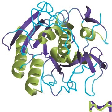
Proteinase K is a stable and highly reactive serine protease. Evidence from crystal and molecular structure studies indicates the enzyme belongs to the subtilisin family with an active-site catalytic triad (Asp39-His69-Ser224). It is stable in a broad range of environments: pH, buffer salts, detergents (SDS), and temperature. In the presence of 0.1-0.5% SDS, proteinase K retains activity and will digest a variety of proteins and nucleases in DNA preparations without compromising the integrity of the isolated DNA.

Unit Definition

One unit will hydrolyze urea-denatured hemoglobin to produce color equivalent to 1.0 μmole of tyrosine per min at pH 7.5 at 37 °C (color by Folin-Ciocalteu reagent).

also commonly purchased with this product

Product No.
Description
Pricing

Pictograms

Exclamation markHealth hazard

Signal Word

Danger

Hazard Statements

Hazard Classifications

Eye Irrit. 2 - Resp. Sens. 1 - Skin Irrit. 2 - STOT SE 3

Target Organs

Respiratory system

Storage Class Code

11 - Combustible Solids

WGK

WGK 1

Flash Point(F)

Not applicable

Flash Point(C)

Not applicable

Personal Protective Equipment

dust mask type N95 (US), Eyeshields, Gloves

Certificates of Analysis (COA)

Search for Certificates of Analysis (COA) by entering the products Lot/Batch Number. Lot and Batch Numbers can be found on a product’s label following the words ‘Lot’ or ‘Batch’.

Already Own This Product?

Find documentation for the products that you have recently purchased in the Document Library.

Visit the Document Library

M M Kristjánsson et al.
European journal of biochemistry, 260(3), 752-760 (1999-04-02)
An extracellular serine proteinase purified from cultures of a psychrotrophic Vibrio species (strain PA-44) belongs to the proteinase K family of the superfamily of subtilisin-like proteinases. The enzyme is secreted as a 47-kDa protein, but under mild heat treatment (30
Rongzhu Cheng et al.
The Journal of biological chemistry, 279(44), 45441-45449 (2004-08-19)
We report here the isolation of a novel acid-labile yellow chromophore from the enzymatic digest of human lens proteins and the identification of its chemical structure by liquid chromatography-mass spectrometry, liquid chromatography-tandem mass spectrometry, and (1)H, (13)C, and two-dimensional NMR.
Nathaniel Denkers et al.
Developmental dynamics : an official publication of the American Association of Anatomists, 229(3), 651-657 (2004-03-03)
Multi-color whole-mount in situ hybridization is a powerful technique for comparing the spatial expression patterns of two or more genes in developing embryos. We have developed an amplified triple-label whole-mount fluorescence in situ hybridization (FISH) protocol that permits detection of
Michela Candelma et al.
Reproduction (Cambridge, England), 153(2), 123-132 (2016-11-03)
In vertebrates, the regulation of gametogenesis is under the control of gonadotropins (Gth), follicle-stimulating hormone (Fsh) and luteinizing hormone (Lh). In fish, the physiological role of Gths is not fully understood, especially in species with asynchronous ovarian development. To elucidate
Janus S Jakobsen et al.
Science advances, 5(7), eaaw4304-eaaw4304 (2019-07-17)
The key myeloid transcription factor (TF), CEBPA, is frequently mutated in acute myeloid leukemia (AML), but the direct molecular effects of this leukemic driver mutation remain elusive. To investigate CEBPA mutant AML, we performed microscale, in vivo chromatin immunoprecipitation sequencing

Articles

Proteinase K (EC 3.4.21.64) activity can be measured spectrophotometrically using hemoglobin as the substrate. Proteinase K hydrolyzes hemoglobin denatured with urea, and liberates Folin-postive amino acids and peptides. One unit will hydrolyze hemoglobin to produce color equivalent to 1.0 μmol of tyrosine per minute at pH 7.5 at 37 °C (color by Folin & Ciocalteu's Phenol Reagent).

Protocols

Proteinase K (EC 3.4.21.64) activity can be measured spectrophotometrically using hemoglobin as the substrate. Proteinase K hydrolyzes hemoglobin denatured with urea, and liberates Folin-postive amino acids and peptides. One unit will hydrolyze hemoglobin to produce color equivalent to 1.0 μmol of tyrosine per minute at pH 7.5 at 37 °C (color by Folin & Ciocalteu's Phenol Reagent).

In Situ Hybridization of Whole-Mount Mouse Embryos with RNA Probes: Hybridization, Washes, and Histochemistry. This is a protocol describing how to perform in situ hybridization on whole mouse embryos. Here we describe the hybridization procedure, and the localization of the DIG-labeled RNA using a conjugate of anti-DIG Fab antibody and calf intestinal alkaline phosphatase. Enzyme activity of the reporter is detected by a color reaction, resulting in the formation of a water-insoluble purple/blue precipitate. Manipulating the Mouse Embryo - Third Edition

Our team of scientists has experience in all areas of research including Life Science, Material Science, Chemical Synthesis, Chromatography, Analytical and many others.

Contact Technical Service