Saltar al contenido
Merck
Todas las fotos(1)

Documentos clave

SRP8056

Sigma-Aldrich

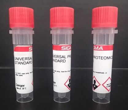
Tim-3 (mouse): FC (human)

recombinant, expressed in CHO cells, ≥98% (SDS-PAGE)

Sinónimos:

HAVcr-2, T-cell immunoglobulin and mucin domain-containing protein 3, TIM3, TIMD3

Iniciar sesiónpara Ver la Fijación de precios por contrato y de la organización


About This Item

Código UNSPSC:
12352200
NACRES:
NA.32

origen biológico

mouse

recombinante

expressed in CHO cells

Ensayo

≥98% (SDS-PAGE)

Formulario

lyophilized

mol peso

monomer 45 kDa by calculation

envase

pkg of 100 μg

condiciones de almacenamiento

avoid repeated freeze/thaw cycles

impurezas

<0.06 EU/μg endotoxin, tested

color

white

Nº de acceso UniProt

Condiciones de envío

wet ice

temp. de almacenamiento

−20°C

Información sobre el gen

mouse ... Havcr2(171285)

Descripción general

Tim-3 is a type I transmembrane protein. It contains an immunoglobulin and a mucin-like domain in its extracellular portion and a tyrosine phosphorylation motif in its cytoplasmic portion. TIM-3 is preferentially expressed on Type 1 T-helper (Th1) and Type 1 CD8+ T (Tc1) cells. TIM-3 is also expressed on some dendritic cells. The gene encoding this protein is localized on mouse chromosome 11B1.1.

Acciones bioquímicas o fisiológicas

The T cell/transmembrane, immunoglobulin and mucin (TIM) family plays a crucial role in regulating immune responses, such as allergy, asthma, transplant tolerance, autoimmunity and also has a control over the response to viral infections. The unique structure of TIM immunoglobulin variable region domains allows highly specific recognition of phosphatidylserine (PtdSer), exposed on the surface of apoptotic cells. Tim-3 generates an inhibitory signal resulting in apoptosis of Type 1 T-helper (Th1) and Type 1 CD8+ T (Tc1) cells. It can also mediate phagocytosis of apoptotic cells and cross-presentation of antigen. It functions to inhibit aggressive Th1-mediated auto- and allo-immune responses. Tim-3 pathway blockade by administration of Tim-3: Fc fusion protein accelerates diabetes in non-obese diabetic mice, causes hyperproliferation of Th1 cells and Th1 cytokine release in an experimental autoimmune encephalomyelitis (EAE) model and prevents acquisition of transplantation tolerance induced by costimulation blockade.

Forma física

Lyophilized from 0.2 μm-filtered solution in PBS.

Reconstitución

Reconstitute at 100 μg/mL in sterile PBS.

Otras notas

The extracellular domain of mouse Tim-3 (aa 21-189) is fused to the N-terminus of the Fc region of human IgG1.

Código de clase de almacenamiento

10 - Combustible liquids

Clase de riesgo para el agua (WGK)

WGK 2

Punto de inflamabilidad (°F)

Not applicable

Punto de inflamabilidad (°C)

Not applicable


Elija entre una de las versiones más recientes:

Certificados de análisis (COA)

Lot/Batch Number

¿No ve la versión correcta?

Si necesita una versión concreta, puede buscar un certificado específico por el número de lote.

¿Ya tiene este producto?

Encuentre la documentación para los productos que ha comprado recientemente en la Biblioteca de documentos.

Visite la Librería de documentos

TIM-3 is Expressed on Activated Human CD4+ T Cells and Regulates Th1 and Th17 Cytokines
William D. Hastings
European Journal of Immunology, 39(9), 2492-2501 (2009)
TIM Proteins and Immunity
Lawrence P. Kane
Journal of Immunology, 184(6), 2743-2749 (2010)
T and B cell hyperactivity and autoimmunity associated with niche-specific defects in apoptotic body clearance in TIM-4-deficient mice
Roselyn R
Proceedings of the National Academy of Sciences of the USA, 107.19, 8706-8711 (2010)
TIM-1 and TIM-3 Proteins in Immune Regulation
Ee Wern Su
Cytokine, 44(1), 9-13 (2008)
TIM genes: a family of cell surface phosphatidylserine receptors that regulate innate and adaptive immunity
Gordon J. Freeman
Immunological Reviews, 235(1), 172-189 (2010)

Nuestro equipo de científicos tiene experiencia en todas las áreas de investigación: Ciencias de la vida, Ciencia de los materiales, Síntesis química, Cromatografía, Analítica y muchas otras.

Póngase en contacto con el Servicio técnico