Saltar al contenido
Merck

C5867

Sigma-Aldrich

Anti-Ciliated Cell Marker antibody, Mouse monoclonal

clone LhS 28, purified from hybridoma cell culture

Iniciar sesiónpara Ver la Fijación de precios por contrato y de la organización


About This Item

Número MDL:
Código UNSPSC:
12352203
NACRES:
NA.41

origen biológico

mouse

Nivel de calidad

conjugado

unconjugated

forma del anticuerpo

purified immunoglobulin

tipo de anticuerpo

primary antibodies

clon

LhS 28, monoclonal

Formulario

buffered aqueous solution

mol peso

antigen ~45 kDa

reactividad de especies

human, hamster

concentración

~2 mg/mL

técnicas

electron microscopy: suitable
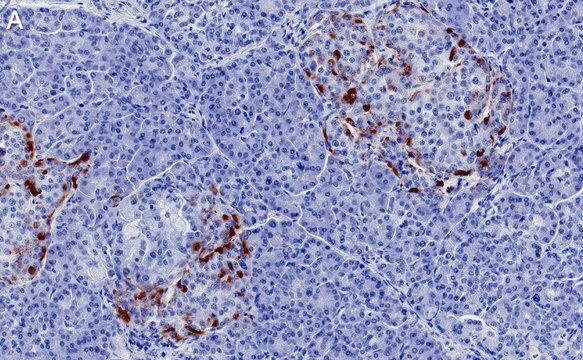
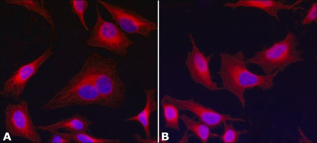
immunocytochemistry: 1-2 μg/mL using BHK a21 cells
immunohistochemistry: suitable
microarray: suitable
western blot: suitable

isotipo

IgG1

Condiciones de envío

dry ice

temp. de almacenamiento

−20°C

modificación del objetivo postraduccional

unmodified

Descripción general

Anti-Ciliated Cell Marker antibody, Mouse monoclonal (mouse IgG1 isotype) is derived from the hybridoma LhS 28 produced by the fusion of mouse myeloma cells (Sp2/0 cells) and splenocytes from BALB/c mice.

Especificidad

Anti-Ciliated Cell Marker antibody, Mouse monoclonal recognizes an antigen in human and hamster ciliated cells.

Inmunógeno

BHK a21 cytoskeletal preparation.

Aplicación

Anti-Ciliated Cell Marker antibody, Mouse monoclonal may be used in:
  • immunoelectron microscopy
  • immunoblotting
  • immunohistochemistry
  • immunocytochemistry

May be used in the detection of ciliated cells and for investigating mechanisms of ciliogenesis.

Acciones bioquímicas o fisiológicas

The mouse hybridoma LhS 28 secretes a monoclonal antibody that recognizes the antigen 44-45 kDa protein associated with the ciliary basal bodies. The antigen recognized by LhS 28 is present in the 9+0 microtubule arrays in the basal body structure of the cilium. It serves as an unequivocal marker for in situ detection and in vitro studies of human ciliated epithelial cells. LhS28 is a potential marker for tubal metaplasia identification and may be useful in recognizing low grade cervical glandular intraepithelial neoplasia. It is effective in detecting ciliogenesis and ciliated cells associated with normal and pathological human specimens

Forma física

Solution in 0.01 M phosphate buffered saline, pH 7.4, containing 15 mM sodium azide.

Almacenamiento y estabilidad

For continuous use, store at 2–8 °C for up to one month. For extended storage, freeze in working aliquots. Repeated freezing and thawing, or storage in “frost-free” freezers, is not recommended. If slight turbidity occurs upon prolonged storage, clarify the solution by centrifugation before use. Working dilution samples should be discarded if not used within 12 hours.

Cláusula de descargo de responsabilidad

Unless otherwise stated in our catalog or other company documentation accompanying the product(s), our products are intended for research use only and are not to be used for any other purpose, which includes but is not limited to, unauthorized commercial uses, in vitro diagnostic uses, ex vivo or in vivo therapeutic uses or any type of consumption or application to humans or animals.

¿No encuentra el producto adecuado?  

Pruebe nuestro Herramienta de selección de productos.

Código de clase de almacenamiento

10 - Combustible liquids

Clase de riesgo para el agua (WGK)

nwg

Punto de inflamabilidad (°F)

Not applicable

Punto de inflamabilidad (°C)

Not applicable


Elija entre una de las versiones más recientes:

Certificados de análisis (COA)

Lot/Batch Number

¿No ve la versión correcta?

Si necesita una versión concreta, puede buscar un certificado específico por el número de lote.

¿Ya tiene este producto?

Encuentre la documentación para los productos que ha comprado recientemente en la Biblioteca de documentos.

Visite la Librería de documentos

M T Comer et al.
Journal of clinical pathology, 52(5), 355-357 (1999-11-24)
The assessment of neoplastic disease in gynaecological histopathology can be complicated by the high incidence of metaplasia seen in tissues of the female genital tract. There is a need to identify specific tissue markers which can be applied in routine

Nuestro equipo de científicos tiene experiencia en todas las áreas de investigación: Ciencias de la vida, Ciencia de los materiales, Síntesis química, Cromatografía, Analítica y muchas otras.

Póngase en contacto con el Servicio técnico