Saltar al contenido
Merck

C2181

Sigma-Aldrich

Anti-Mouse IgG (whole molecule) F(ab′)2 fragment–Cy3 antibody produced in sheep

affinity isolated antibody, buffered aqueous solution

Sinónimos:

Cy3 Anti-Mouse IgG, Cy3 Mouse IgG, Sheep Anti-Mouse IgG

Iniciar sesiónpara Ver la Fijación de precios por contrato y de la organización


About This Item

Número MDL:
Código UNSPSC:
12352203
NACRES:
NA.46

origen biológico

sheep

Nivel de calidad

conjugado

CY3 conjugate

forma del anticuerpo

affinity isolated antibody

tipo de anticuerpo

secondary antibodies

clon

polyclonal

Formulario

buffered aqueous solution

reactividad de especies

mouse

técnicas

immunohistochemistry (formalin-fixed, paraffin-embedded sections): 1:100

Condiciones de envío

wet ice

temp. de almacenamiento

2-8°C

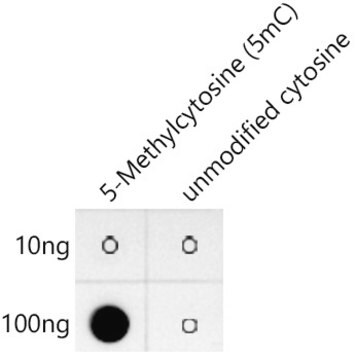
modificación del objetivo postraduccional

unmodified

Descripción general

Immunoglobulins (Igs) belong to the immunoglobulin super-family. IgG is an abundant protein in human serum. The four classes of IgG include IgG1, IgG2, IgG3 and IgG4.. The IgG heavy chain region is mapped to human chromosome 14. Igs have two heavy (H) and two light (L) chains, held together by disulfide linkages. The heavy chain has one variable N-terminal region and three to four constant (CH1-CH4) C-terminal regions. The L chain comprises of one variable N-terminal region and a constant C-terminal region.

Aplicación

Anti-Mouse IgG (whole molecule) F(ab′)2 fragment-Cy3 antibody produced in sheep was used for BrdU staining of frozen quail muscle cross sections at a dilution of 1:200 to analyze activated, proliferating muscle precursor cells.
Anti-Mouse IgG (whole molecule) F(ab′)2 fragment−Cy3 antibody produced in sheep has been used:
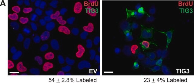
  • in immunolabeling of Hela cells
  • as secondary antibody in immunocytochemistry of dendritic cells
  • as secondary antibody in immunofluorescence analysis of mesenchymal stem cells
  • as secondary antibody in immunofluorescence staining keratinocyte cell lines
  • in immunohistochemistry of articular cartilage

The product binds to all mouse Igs and is useful when trying to avoid background staining due to the presence of Fc receptors.

Acciones bioquímicas o fisiológicas

Digestion of IgG by papain results in the generation of fragment antigen binding (Fab). Pepsin digestion of IgG results in fragment crystallizable (Fc). The Fc region of IgG antibody has enormous therapeutic potential and is exploited for the development of therapeutic antibodies.
IgG antibody subtype is the most abundant of serum immunoglobulins of the immune system. It is secreted by B cells and is found in blood and extracellular fluids and provides protection from infections caused by bacteria, fungi and viruses. Maternal IgG is transferred to fetus through the placenta that is vital for immune defense of the neonate against infections. The coupling of Cy3 to Anti-Mouse IgG (whole molecule) F(ab′)2 fragment antibody allows for the visualization of protein by fluorescent microscopy.

Otras notas

Antibody adsorbed with human serum proteins.

Forma física

Solution in 0.01 M phosphate buffered saline, pH 7.4, containing 1% bovine serum albumin and 15 mM sodium azide.

Nota de preparación

Adsorbed to reduce background staining with human samples.

Cláusula de descargo de responsabilidad

Unless otherwise stated in our catalog or other company documentation accompanying the product(s), our products are intended for research use only and are not to be used for any other purpose, which includes but is not limited to, unauthorized commercial uses, in vitro diagnostic uses, ex vivo or in vivo therapeutic uses or any type of consumption or application to humans or animals.

¿No encuentra el producto adecuado?  

Pruebe nuestro Herramienta de selección de productos.

Código de clase de almacenamiento

10 - Combustible liquids

Clase de riesgo para el agua (WGK)

nwg

Punto de inflamabilidad (°F)

Not applicable

Punto de inflamabilidad (°C)

Not applicable


Elija entre una de las versiones más recientes:

Certificados de análisis (COA)

Lot/Batch Number

¿No ve la versión correcta?

Si necesita una versión concreta, puede buscar un certificado específico por el número de lote.

¿Ya tiene este producto?

Encuentre la documentación para los productos que ha comprado recientemente en la Biblioteca de documentos.

Visite la Librería de documentos

Identification of a Novel Keratin 9 Missense Mutation in a Chinese Family with Epidermolytic Palmoplantar Keratoderma.
Xiao H, et al.
Cellular Physiology and Biochemistry, 46(5), 1919-1929 (2018)
A Birkmann et al.
Journal of virology, 75(23), 11583-11593 (2001-11-02)
An immunodominant envelope glycoprotein is encoded by the human herpesvirus 8 (HHV-8) (also termed Kaposi's sarcoma-associated herpesvirus) K8.1 gene. The functional role of glycoprotein K8.1 is unknown, and recognizable sequence homology to K8.1 is not detectable in the genomes of
Agili-C implant promotes the regenerative capacity of articular cartilage defects in an ex vivo model.
Chubinskaya S, et al.
Knee Surgery, Sports Traumatology, Arthroscopy : Official Journal of the ESSKA, 46(5), 1-12 (2018)
Antonio F Ribeiro et al.
Scientific reports, 9(1), 11842-11842 (2019-08-16)
Satellite cells (SCs) are the main muscle stem cells responsible for its regenerative capacity. In muscular dystrophies, however, a failure of the regenerative process results in muscle degeneration and weakness. To analyze the effect of different degrees of muscle degeneration
Visualization of altered replication dynamics after DNA damage in human cells.
Merrick C, et al.
The Journal of Biological Chemistry, 279(19), 20067-20075 (2004)

Nuestro equipo de científicos tiene experiencia en todas las áreas de investigación: Ciencias de la vida, Ciencia de los materiales, Síntesis química, Cromatografía, Analítica y muchas otras.

Póngase en contacto con el Servicio técnico