93606
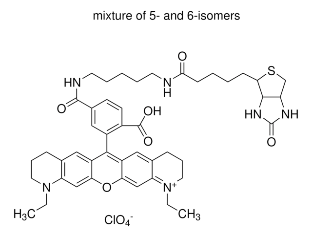
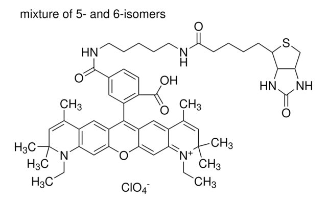
Atto 647N-Biotin
BioReagent, suitable for fluorescence, ≥90.0% (HPLC)
Sinónimos:
Biotin-Atto 647N
Iniciar sesiónpara Ver la Fijación de precios por contrato y de la organización
About This Item
Código UNSPSC:
12161900
NACRES:
NA.25
Productos recomendados
Línea del producto
BioReagent
Análisis
≥90.0% (HPLC)
formulario
powder
fabricante / nombre comercial
ATTO-TEC GmbH
λ
in ethanol (with 0.1% trifluoroacetic acid)
Absorción UV
λ: 642.0-648.0 nm Amax
idoneidad
suitable for fluorescence
temp. de almacenamiento
−20°C
Aplicación
Atto fluorescent labels are designed for high sensitivity applications, including single molecule detection. Atto labels have rigid structures that do not show any cis-trans-isomerization. Thus these labels display exceptional intensity with minimal spectral shift on conjugation.
Código de clase de almacenamiento
11 - Combustible Solids
Clase de riesgo para el agua (WGK)
WGK 3
Punto de inflamabilidad (°F)
Not applicable
Punto de inflamabilidad (°C)
Not applicable
Equipo de protección personal
Eyeshields, Gloves, type N95 (US)
Certificados de análisis (COA)
Busque Certificados de análisis (COA) introduciendo el número de lote del producto. Los números de lote se encuentran en la etiqueta del producto después de las palabras «Lot» o «Batch»
¿Ya tiene este producto?
Encuentre la documentación para los productos que ha comprado recientemente en la Biblioteca de documentos.
Los clientes también vieron
STED microscopy to monitor agglomeration of silica particles inside A549 cells.
Schubbe, S., et al.
Advanced Engineering Materials, 12, 417-422 (2010)
A novel nanoscopic tool by combining AFM with STED microscopy.
Harke, B., et al.
Optical Nanoscopy, 1, 3-3 (2012)
Volker Westphal et al.
Science (New York, N.Y.), 320(5873), 246-249 (2008-02-23)
We present video-rate (28 frames per second) far-field optical imaging with a focal spot size of 62 nanometers in living cells. Fluorescently labeled synaptic vesicles inside the axons of cultured neurons were recorded with stimulated emission depletion (STED) microscopy in
Marisa L Martin-Fernandez et al.
International journal of molecular sciences, 13(11), 14742-14765 (2012-12-04)
Insights from single-molecule tracking in mammalian cells have the potential to greatly contribute to our understanding of the dynamic behavior of many protein families and networks which are key therapeutic targets of the pharmaceutical industry. This is particularly so at
S E D Webb et al.
Optics express, 16(25), 20258-20265 (2008-12-10)
We combine single molecule fluorescence orientation imaging with single-pair fluorescence resonance energy transfer microscopy, using a total internal reflection microscope. We show how angles and FRET efficiencies can be determined for membrane proteins at the single molecule level and provide
Nuestro equipo de científicos tiene experiencia en todas las áreas de investigación: Ciencias de la vida, Ciencia de los materiales, Síntesis química, Cromatografía, Analítica y muchas otras.
Póngase en contacto con el Servicio técnico


