
MABN1793
Anti-SorLA Antibody, clone 20C11
clone 20C11, from mouse
Sinónimos:
Sortilin-related receptor, Gp250, LDLR relative with 11 ligand-binding repeats, Low-density lipoprotein receptor relative with 11 ligand-binding repeats, LR11, SorLA, SorLA-1, Sorting protein-related receptor containing LDLR class A repeats
About This Item
Productos recomendados


origen biológico
mouse
Nivel de calidad
forma del anticuerpo
purified immunoglobulin
tipo de anticuerpo
primary antibodies
clon
20C11, monoclonal
reactividad de especies
mouse, human
técnicas
ELISA: suitable
immunocytochemistry: suitable
western blot: suitable
isotipo
IgG1κ
Nº de acceso NCBI
Nº de acceso UniProt
modificación del objetivo postraduccional
unmodified
Información sobre el gen
human ... SORL1(6653)
Descripción general
Especificidad
Inmunógeno
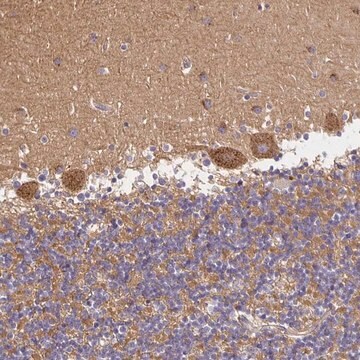
Aplicación
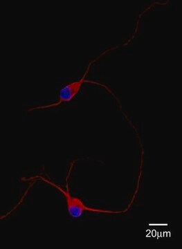
Immunocytochemistry Analysis: A representative lot detected SorLA immunoreactivity primarily located at the cell body (soma) by fluorescent immunocytochemistry staining of 4% paraformaldehyde-fixed, 0.1% Triton X-100-permeabilized primary murine hippocampal neurons (Gustafsen, C., et al. (2013). J. Neurosci. 33(1):64-71).
Immunocytochemistry Analysis: A representative lot immunostained endocytic vesicular structures containing endocytosed sAPP by fluorescent immunocytochemistry staining of 4% paraformaldehyde-fixed, 0.1% Triton X-100-permeabilized HEK293 cells expressing exogenously transfected human SorLA (Gustafsen, C., et al. (2013). J. Neurosci. 33(1):64-71).
Immunocytochemistry Analysis: A representative lot detected the cellular localization of exogenously expressed wild-type and the FANSHY-to-6A4 mutant human SorLA by fluorescent immuncytochemistry staining of 4% paraformaldehyde-fixed, 0.5% saponin-permeabilized SH-SY5Y transfectants (Fjorback, A.W., et al. (2012). J. Neurosci. 32(4):1467-1480).
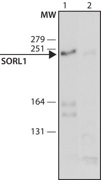
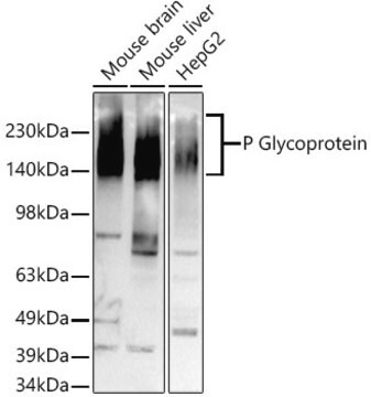
Western Blotting Analysis: A representative lot detected endogenous SorLA in sucrose gradient-fractionated mouse brain extracts as well as the expression of exogenously transfected human SorLA in HEK293 transfectants (Gustafsen, C., et al. (2013). J. Neurosci. 33(1):64-71).
ELISA Analysis: Representative lots were employed as the detection antibody for quantiating SorLA level in human brain cortical extracts as well as the level of exogenously expressed human SorLA in CHO cells (Caglayan, S., et al. (2012). Arch. Neurol. 69(3):373-379; Schmidt, V., et al. (2012). EMBO J. 31(1):187-200).
Calidad
Immunocytochemistry Analysis: 4.0 µg/mL of this antibody immunostained SorLA endocytic vesicles in HEK293 cells expressing transfected human SorLA.
Descripción de destino
Forma física
Otras notas
¿No encuentra el producto adecuado?
Pruebe nuestro Herramienta de selección de productos.
Código de clase de almacenamiento
12 - Non Combustible Liquids
Clase de riesgo para el agua (WGK)
WGK 1
Punto de inflamabilidad (°F)
Not applicable
Punto de inflamabilidad (°C)
Not applicable
Certificados de análisis (COA)
Busque Certificados de análisis (COA) introduciendo el número de lote del producto. Los números de lote se encuentran en la etiqueta del producto después de las palabras «Lot» o «Batch»
¿Ya tiene este producto?
Encuentre la documentación para los productos que ha comprado recientemente en la Biblioteca de documentos.
Nuestro equipo de científicos tiene experiencia en todas las áreas de investigación: Ciencias de la vida, Ciencia de los materiales, Síntesis química, Cromatografía, Analítica y muchas otras.
Póngase en contacto con el Servicio técnico