MAB329-C
Anti-Synaptophysin Antibody, clone SP15 (Ascites Free)
clone SP15, from mouse
Sinónimos:
Major synaptic vesicle protein p38, Synaptophysin
About This Item
Productos recomendados
origen biológico
mouse
Nivel de calidad
forma del anticuerpo
purified antibody
tipo de anticuerpo
primary antibodies
clon
SP15, monoclonal
reactividad de especies
human, monkey, feline, rat, mouse
técnicas
ELISA: suitable
immunocytochemistry: suitable
immunohistochemistry: suitable
western blot: suitable
isotipo
IgMκ
Nº de acceso NCBI
Nº de acceso UniProt
modificación del objetivo postraduccional
unmodified
Información sobre el gen
human ... SYP(6855)
Descripción general
Inmunógeno
Aplicación
Neuroscience
Developmental Neuroscience
Western Blotting Analysis: A representative lot detected synaptophysin in rat hippocampal tissue extracts (Lin, D., et al. (2012). Behav. Brain Res. 228(2):319-327).
Western Blotting Analysis: Representative lots detected synaptophysin in mouse brain synaptosomes preparations (Shim, S.Y., et al. (2008). J. Neurosci. 28(14):3604-3614).
Western Blotting Analysis: A representative lot detected synaptophysin in the large dense-core vesicles-containing fractions obtained by sucrose gradient centrifugation of rat brain median eminence (ME) neuroterminals preparations (Yin, W., et al. (2007). Exp. Biol. Med. (Maywood). 232(5):662-673).
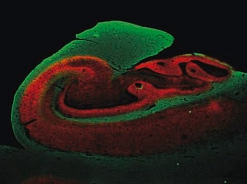
Immunohistochemistry Analysis: A representative lot detected a stronger synaptophysin immunoreactivity in the inner molecular layer of frozen dentate gyrus sections from a 3-week old monkey, while a stronger immunoreactivity was seen in the outer molecular layer of dentate gyrus sections from a 13-year old monkey (Lavenex, P., et al. (2007). Dev. Neurosci. 29(1-2):179–192).
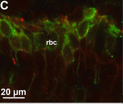
Immunohistochemistry Analysis: A representative lot immunostained nerve terminals at palisade ending using cat extraocular muscle (EOM) whole mount sections (Konakci, K.Z., et al. (2005). Invest. Ophthalmol. Vis. Sci. 46(1):155-165).
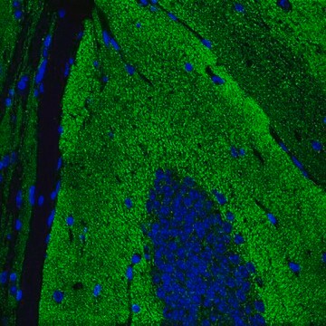
Immunohistochemistry Analysis: A representative lot detected synaptophysin immunoreactivity in human brain tissue sections (Honer, W.G., et al. (1993). Brain Res. 609(1-2):9-20).
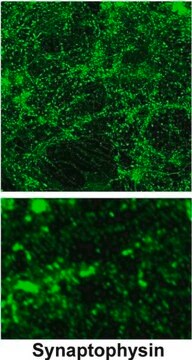
Immunocytochemistry Analysis: A representative lot detected punctate synatophysin immunoreactivity among cultured primary cerebrocortical neurons from rat embryos (Bragina, L., et al. (2006). J. Neurochem. 99(1):134-141.).
ELISA Analysis: Representative lots were employed as the capture antibody for the detection of synaptophysin in human temporal cortex, frontal cortex, and cerebellar cortex extracts by sandwich ELISA (Klucken, J., et al. (2006). Acta Neuropathol. 111(2):101-108; Fukumoto, H., et al. (2002). Arch. Neurol. 59(9):1381-1389).
Calidad
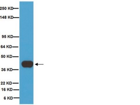
Western Blotting Analysis: 1.0 µg/mL of this antibody detected Synaptophysin in 10 µg of rat brain tissue lysate.
Descripción de destino
Forma física
Almacenamiento y estabilidad
Otras notas
Cláusula de descargo de responsabilidad
¿No encuentra el producto adecuado?
Pruebe nuestro Herramienta de selección de productos.
Opcional
Código de clase de almacenamiento
12 - Non Combustible Liquids
Clase de riesgo para el agua (WGK)
WGK 2
Punto de inflamabilidad (°F)
Not applicable
Punto de inflamabilidad (°C)
Not applicable
Certificados de análisis (COA)
Busque Certificados de análisis (COA) introduciendo el número de lote del producto. Los números de lote se encuentran en la etiqueta del producto después de las palabras «Lot» o «Batch»
¿Ya tiene este producto?
Encuentre la documentación para los productos que ha comprado recientemente en la Biblioteca de documentos.
Nuestro equipo de científicos tiene experiencia en todas las áreas de investigación: Ciencias de la vida, Ciencia de los materiales, Síntesis química, Cromatografía, Analítica y muchas otras.
Póngase en contacto con el Servicio técnico