Saltar al contenido
Merck
Todas las fotos(2)

Key Documents

ABS2269

Sigma-Aldrich

Anti-TRPM4

Sinónimos:

Transient receptor potential cation channel subfamily M member 4;Calcium-activated non-selective cation channel 1;Long transient receptor potential channel 4;TrpC-4;LTrpC4;MLS2s;Melastatin-like 2

Iniciar sesiónpara Ver la Fijación de precios por contrato y de la organización


About This Item

Código UNSPSC:
12352203
NACRES:
NA.43

origen biológico

rabbit

Nivel de calidad

forma del anticuerpo

purified antibody

tipo de anticuerpo

primary antibodies

mol peso

calculated mol wt 135.34 kDa
observed mol wt ~135 kDa

purificado por

affinity chromatography

reactividad de especies

rat, mouse

envase

antibody small pack of 100 μg

técnicas

immunocytochemistry: suitable
immunohistochemistry: suitable
western blot: suitable

isotipo

IgG

secuencia del epítopo

C-terminal extracellular half

Nº de acceso Protein ID

Nº de acceso UniProt

temp. de almacenamiento

-10 to -25°C

modificación del objetivo postraduccional

unmodified

Información sobre el gen

Descripción general

Transient receptor potential cation channel subfamily M member 4 (UniProt: Q9ESQ5; also known as Calcium-activated non-selective cation channel 1, Long transient receptor potential channel 4, LTrpC-4, LTrpC4, MLS2s, Melastatin-like 2) is encoded by the Trpm4 (also known as Ltrpc4) gene (Gene ID: 171143) in rat. TRPM4 is a multi-pass membrane protein with four cytoplasmic domains; six transmembrane domains; and four extracellular domains. Two isoforms of TRPM4 have been described that are produced by alternative splicing. Isoform 1 is highly expressed in the testis tissue with a moderate expression in brain, spleen, and thymus. Isoform 2 is expressed in the brain and spleen. It serves as a calcium-activated non-selective cation channel that mediates membrane depolarization. Although it is a calcium activated channel, it is impermeable to calcium. It mediates the transport of monovalent cations that leads to membrane depolarization. It is reported to participates in T-cell activation by modulating Ca2+ oscillations after T lymphocyte activation, which is required for NFAT-dependent IL-2 production. TRPM4 is also involved in myogenic constriction of cerebral arteries. It plays a role in control of insulin secretion in pancreatic β-cells. It may also be involved in pacemaking activity or could cause irregular electrical activity under conditions of Calcium overload. It is known to affect Th1 and Th2 cell motility and cytokine production through differential regulation of calcium signaling and NFATC1 localization. This polyclonal antibody is reported toblock the channel vua binding to a region close to the channel porefrom extracellular space. Bu blocking TRPM4, it can downregulate TRPM4 surface expression and prevent hypoxia-induced cell swelling. (Ref.: Wei, S., et al. (2020). Front Cell Dev Bio. 8; 562584; Chen, B;et al. (2019). Pflugers Arch. 471(11-12); 1455-1466; Guinamard, R., et al. (2006). Hypertension. 48(4); 587-594; Launay, P., et al. (2002). Cell. 109(3); 397-407).

Especificidad

This rabbit polyclonal antibody detects Transient receptor potential cation channel subfamily M member 4 (TRPM4). It targets an epitope within 24 amino acids from the C-terminal, extracellular domain.

Inmunógeno

GST-conjugated linear peptide corresponding to 28 amino acids between transmembrane segment 5 and 6 from the C-terminal half of rat TRPM4.

Aplicación

Quality Control Testing

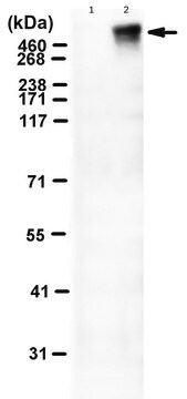
Evaluated by Western Blotting in lysate from HEK293 cells transfected with Mouse TRPM4.

Western Blotting Analysis (WB): A 1:1,000 dilution of this antibody detected TRPM4 in lysate from HEK293 cells transfected with Mouse TRPM4.

Tested Applications

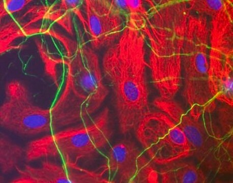
Immunocytochemistry Analysis: A representative lot detected TRPM4 in Immunocytochemistry application (Low, S.W., et. al. (2021). Sci Rep. 11(1):10411).

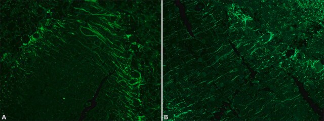
Immunohistochemistry (Paraffin) Analysis: A 1:250 dilution from a representative lot detected TRPM4 in rat spinal cord tissue sections.

Note: Actual optimal working dilutions must be determined by end user as specimens, and experimental conditions may vary with the end user.

Forma física

Purified rabbit polyclonal antibody in PBS without azide.

Reconstitución

1.0 mg/mL. Please refer to guidance on suggested starting dilutions and/or titers per application and sample type.

Almacenamiento y estabilidad

Store at -10°C to -25°C. Handling Recommendations: Upon receipt and prior to removing the cap, centrifuge the vial and gently mix the solution. Aliquot into microcentrifuge tubes and store at -20°C. Avoid repeated freeze/thaw cycles, which may damage IgG and affect product performance.

Otras notas

Concentration: Please refer to the Certificate of Analysis for the lot-specific concentration.

Cláusula de descargo de responsabilidad

Unless otherwise stated in our catalog or other company documentation accompanying the product(s), our products are intended for research use only and are not to be used for any other purpose, which includes but is not limited to, unauthorized commercial uses, in vitro diagnostic uses, ex vivo or in vivo therapeutic uses or any type of consumption or application to humans or animals.

¿No encuentra el producto adecuado?  

Pruebe nuestro Herramienta de selección de productos.

Código de clase de almacenamiento

12 - Non Combustible Liquids

Clase de riesgo para el agua (WGK)

WGK 2

Punto de inflamabilidad (°F)

Not applicable

Punto de inflamabilidad (°C)

Not applicable


Certificados de análisis (COA)

Busque Certificados de análisis (COA) introduciendo el número de lote del producto. Los números de lote se encuentran en la etiqueta del producto después de las palabras «Lot» o «Batch»

¿Ya tiene este producto?

Encuentre la documentación para los productos que ha comprado recientemente en la Biblioteca de documentos.

Visite la Librería de documentos

Nuestro equipo de científicos tiene experiencia en todas las áreas de investigación: Ciencias de la vida, Ciencia de los materiales, Síntesis química, Cromatografía, Analítica y muchas otras.

Póngase en contacto con el Servicio técnico