Saltar al contenido
Merck
Todas las fotos(2)

Documentos clave

AB6017

Sigma-Aldrich

Anti-F-actin-capping protein subunit beta Antibody

from rabbit

Sinónimos:

capping protein (actin filament) muscle Z-line, beta, F-actin capping protein beta subunit

Iniciar sesiónpara Ver la Fijación de precios por contrato y de la organización


About This Item

Código UNSPSC:
12352203
eCl@ss:
32160702
NACRES:
NA.41

origen biológico

rabbit

Nivel de calidad

forma del anticuerpo

purified antibody

tipo de anticuerpo

primary antibodies

clon

polyclonal

reactividad de especies

mouse, rat, human

reactividad de especies (predicha por homología)

canine (based on 100% sequence homology), primate (based on 100% sequence homology), bovine (based on 100% sequence homology)

técnicas

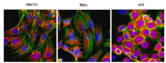
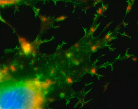
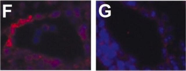
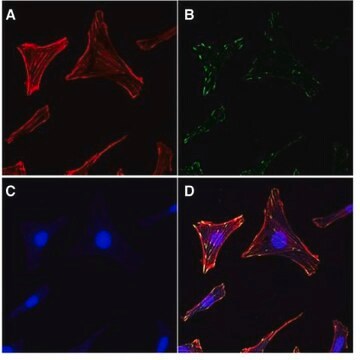
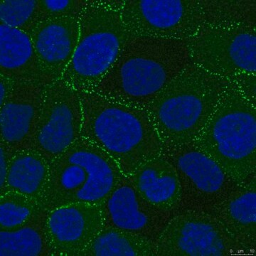
immunocytochemistry: suitable
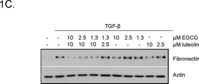
western blot: suitable

Nº de acceso NCBI

Nº de acceso UniProt

Condiciones de envío

wet ice

modificación del objetivo postraduccional

unmodified

Información sobre el gen

bovine ... Capzb(338052)
dog ... Capzb(478209)
human ... CAPZB(832)
mouse ... Capzb(12345)
rat ... Capzb(298584)

Descripción general

F-actin-capping-protein subunit beta (CapZ beta) belongs to the F-actin capping protein family, which are characteristic heterodimers, consisting of an α subunit (31-36 kDa) and a β subunit (28-32 kDa), that cap the barbed end of actin filaments within all eukaryotes. Their ability to bind the actin filaments is in a manner independent of Ca2++ and requires the C-terminal end of both subunits for optimal binding. F-actin capping protein subunit beta is contained within the Z-discs of striated muscle, where it functions to inhibit the polymerization and depolymerization of actin. Within vertebrates, there are three isoforms for each of F-actin capping protein subunit beta’s subunits. While there is almost nothing known regarding the β3 subunit, the β1 and β2 isoforms each have distinct roles. The isoform β2 is predominantly expressed in non-muscle cells and is found in intercalated discs and the cell periphery, but is not observed at Z-discs. Isoform β1 is more greatly expressed in striated muscle and is localized at the Z-discs. It also contains the COOH-terminal extension necessary for capping the actin.

Especificidad

The antibody recognizes F-actin-capping protein subunit beta at the C-terminus of isoform 2.

Inmunógeno

Epitope: C-terminus of Isoform 2
KLH-conjugated linear peptide corresponding to human F-actin-capping protein subunit beta of the C-terminus of isoform 2.

Aplicación

Detect F-actin-capping protein subunit beta using this Anti-F-actin-capping protein subunit beta Antibody validated for use in WB & IC.
Immunocytochemistry Analysis: 1:500 dilution of the antibody has been shown to detect F-actin-capping protein subunit beta in NIH/3T3 and A431 cells.
Research Category
Cell Structure
Research Sub Category
Cytoskeleton

Calidad

Evaluated by Western Blot in HeLa cell lysate.

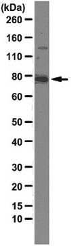
Western Blot Analysis: 0.1 µg/mL of this antibody detected F-actin-capping protein subunit beta in 10 µg of HeLa cell lysate.

Descripción de destino

~ 34 kDa observed MW. There are 3 isoforms produced by alternative splicing: Isoform 1 at 31 kDa, Isoform 2 at 31 kDa, and Isoform 3 (sequence not available).

Forma física

Format: Purified
Protein A purified
Purified rabbit polyclonal in buffer containing 0.1 M Tris-Glycine, pH 7.4, 150 mM NaCl, with 0.05% sodium azide.

Almacenamiento y estabilidad

Stable for 1 year at 2-8°C from date of receipt.

Nota de análisis

Control
HeLa cell lysate

Otras notas

Concentration: Please refer to the Certificate of Analysis for the lot-specific concentration.

Cláusula de descargo de responsabilidad

Unless otherwise stated in our catalog or other company documentation accompanying the product(s), our products are intended for research use only and are not to be used for any other purpose, which includes but is not limited to, unauthorized commercial uses, in vitro diagnostic uses, ex vivo or in vivo therapeutic uses or any type of consumption or application to humans or animals.

¿No encuentra el producto adecuado?  

Pruebe nuestro Herramienta de selección de productos.

Código de clase de almacenamiento

12 - Non Combustible Liquids

Clase de riesgo para el agua (WGK)

WGK 1

Punto de inflamabilidad (°F)

Not applicable

Punto de inflamabilidad (°C)

Not applicable


Certificados de análisis (COA)

Busque Certificados de análisis (COA) introduciendo el número de lote del producto. Los números de lote se encuentran en la etiqueta del producto después de las palabras «Lot» o «Batch»

¿Ya tiene este producto?

Encuentre la documentación para los productos que ha comprado recientemente en la Biblioteca de documentos.

Visite la Librería de documentos

Shamim A Sinnar et al.
Molecular biology of the cell, 25(14), 2152-2160 (2014-05-16)
Capping protein (CP) binds to barbed ends of growing actin filaments and inhibits elongation. CP is essential for actin-based motility in cell-free systems and in Dictyostelium. Even though CP is believed to be critical for creating the lamellipodial actin structure
Ying-Hsi Lin et al.
Journal of muscle research and cell motility, 36(4-5), 329-337 (2015-10-03)
The heart is exquisitely sensitive to mechanical stimuli and adapts to increased demands for work by enlarging the cardiomyocytes. In order to determine links between mechano-transduction mechanisms and hypertrophy, neonatal rat ventricular myocytes (NRVM) were subjected to physiologic strain for
Haibo Du et al.
Frontiers in cell and developmental biology, 9, 765559-765559 (2021-11-09)
Stereocilia are actin-based cell protrusions on the apical surface of inner ear hair cells, playing a pivotal role in hearing and balancing sensation. The development and maintenance of stereocilia is tightly regulated and deficits in this process usually lead to
Keji Yan et al.
Frontiers in molecular neuroscience, 15, 829204-829204 (2022-03-05)
Hair cells are mechanosensitive cells in the inner ear, characterized by dozens to hundreds of actin-based stereocilia and one tubulin-based kinocilium on the apical surface of each cell. Two types of hair cells, namely cochlear hair cells and vestibular hair
Erin F Spence et al.
Nature communications, 10(1), 386-386 (2019-01-25)
Excitatory synapse formation during development involves the complex orchestration of both structural and functional alterations at the postsynapse. However, the molecular mechanisms that underlie excitatory synaptogenesis are only partially resolved, in part because the internal machinery of developing synapses is

Nuestro equipo de científicos tiene experiencia en todas las áreas de investigación: Ciencias de la vida, Ciencia de los materiales, Síntesis química, Cromatografía, Analítica y muchas otras.

Póngase en contacto con el Servicio técnico