Skip to Content
Merck
All Photos(5)

Key Documents

HPA002951

Sigma-Aldrich

Anti-CSPG4 Antibody

enhanced validation

Prestige Antibodies® Powered by Atlas Antibodies, rabbit polyclonal

Synonym(s):

Anti-Chondroitin sulfate proteoglycan 4 precursor, Anti-Chondroitin sulfate proteoglycan NG2, Anti-Melanoma chondroitin sulfate proteoglycan, Anti-Melanoma-associated chondroitin sulfate proteoglycan

Sign Into View Organizational & Contract Pricing


About This Item

UNSPSC Code:
12352203
Human Protein Atlas Number:
NACRES:
NA.41

product name

Anti-CSPG4 antibody produced in rabbit, Prestige Antibodies® Powered by Atlas Antibodies, affinity isolated antibody, buffered aqueous glycerol solution

biological source

rabbit

Quality Level

conjugate

unconjugated

antibody form

affinity isolated antibody

antibody product type

primary antibodies

clone

polyclonal

product line

Prestige Antibodies® Powered by Atlas Antibodies

form

buffered aqueous glycerol solution

species reactivity

human

enhanced validation

recombinant expression
Learn more about Antibody Enhanced Validation

technique(s)

immunoblotting: 0.04-0.4 μg/mL
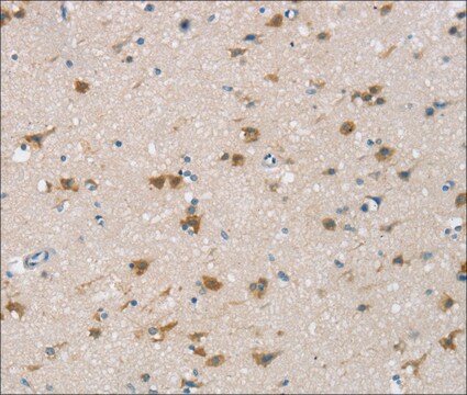
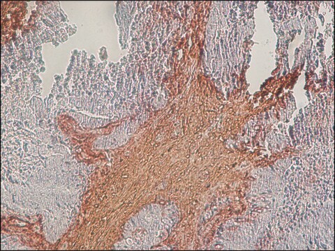
immunohistochemistry: 1:200-1:500

immunogen sequence

ILYEHEMPPEPFWEAHDTLELQLSSPPARDVAATLAVAVSFEAACPQRPSHLWKNKGLWVPEGQRARITVAALDASNLLASVPSPQRSEHDVLFQVTQFPSRGQLLVSEEPLHAGQPHFLQSQLAAGQLVYAHGGGGTQQDGFHFRAHL

UniProt accession no.

application(s)

research pathology

shipped in

wet ice

storage temp.

−20°C

target post-translational modification

unmodified

Gene Information

human ... CSPG4(1464)

General description

Chondroitin sulfate proteoglycan 4 (CSPG4) belongs to the chondroitin sulfate group of glycosaminoglycans (GAGs) and is a type 1 transmembrane protein that harbors glycosylation. The CSPG4 gene is mapped to human chromosome15q24.2. It is an N-linked glycoprotein with a large extracellular domain, transmembrane domain and a cytoplasmic tail. The extracellular domain comprises two laminin G-type subdomains and a middle domain with CSPG4 specific repeats.

Immunogen

Chondroitin sulfate proteoglycan 4 precursor recombinant protein epitope signature tag (PrEST)

Application

All Prestige Antibodies Powered by Atlas Antibodies are developed and validated by the Human Protein Atlas (HPA) project and as a result, are supported by the most extensive characterization in the industry.

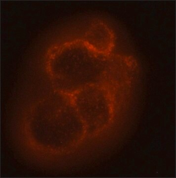
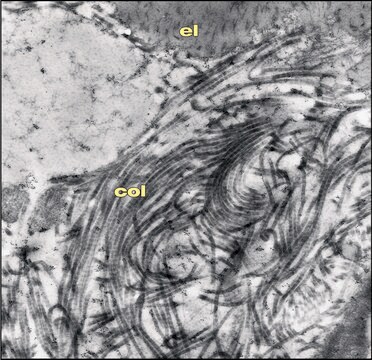
The Human Protein Atlas project can be subdivided into three efforts: Human Tissue Atlas, Cancer Atlas, and Human Cell Atlas. The antibodies that have been generated in support of the Tissue and Cancer Atlas projects have been tested by immunohistochemistry against hundreds of normal and disease tissues and through the recent efforts of the Human Cell Atlas project, many have been characterized by immunofluorescence to map the human proteome not only at the tissue level but now at the subcellular level. These images and the collection of this vast data set can be viewed on the Human Protein Atlas (HPA) site by clicking on the Image Gallery link. We also provide Prestige Antibodies® protocols and other useful information.
Anti-CSPG4 antibody produced in rabbit has been used in fluorescent immunohistochemistry(1:200), 1:250 and immunocytochemistry.

Biochem/physiol Actions

Chondroitin sulfate proteoglycan 4 (CSPG4) is implicated in the pathogenesis of glioma proliferation. It may be involved in the regulation of the neural network, oligodendrocyte formation, and epithelial stem cell homeostasis. Missense mutations in the CSPG4 gene may correlate with white matter integrity impairment and schizophrenia development. It regulates cytoskeletal reorganization and epithelial to mesenchymal transition (EMT) in melanomas. CSPG4 may serve as a potential target for cancer therapies.

Features and Benefits

Prestige Antibodies® are highly characterized and extensively validated antibodies with the added benefit of all available characterization data for each target being accessible via the Human Protein Atlas portal linked just below the product name at the top of this page. The uniqueness and low cross-reactivity of the Prestige Antibodies® to other proteins are due to a thorough selection of antigen regions, affinity purification, and stringent selection. Prestige antigen controls are available for every corresponding Prestige Antibody and can be found in the linkage section.

Every Prestige Antibody is tested in the following ways:
  • IHC tissue array of 44 normal human tissues and 20 of the most common cancer type tissues.
  • Protein array of 364 human recombinant protein fragments.

Linkage

Corresponding Antigen APREST86400

Physical form

Solution in phosphate-buffered saline, pH 7.2, containing 40% glycerol and 0.02% sodium azide

Legal Information

Prestige Antibodies is a registered trademark of Merck KGaA, Darmstadt, Germany

Disclaimer

Unless otherwise stated in our catalog or other company documentation accompanying the product(s), our products are intended for research use only and are not to be used for any other purpose, which includes but is not limited to, unauthorized commercial uses, in vitro diagnostic uses, ex vivo or in vivo therapeutic uses or any type of consumption or application to humans or animals.

Not finding the right product?  

Try our Product Selector Tool.

Storage Class Code

10 - Combustible liquids

WGK

WGK 1

Flash Point(F)

Not applicable

Flash Point(C)

Not applicable

Personal Protective Equipment

dust mask type N95 (US), Eyeshields, Gloves

Certificates of Analysis (COA)

Search for Certificates of Analysis (COA) by entering the products Lot/Batch Number. Lot and Batch Numbers can be found on a product’s label following the words ‘Lot’ or ‘Batch’.

Already Own This Product?

Find documentation for the products that you have recently purchased in the Document Library.

Visit the Document Library

Femke M de Vrij et al.
Molecular psychiatry, 24(5), 757-771 (2018-01-06)
Schizophrenia is highly heritable, yet its underlying pathophysiology remains largely unknown. Among the most well-replicated findings in neurobiological studies of schizophrenia are deficits in myelination and white matter integrity; however, direct etiological genetic and cellular evidence has thus far been
Mikkel G Terp et al.
Cancer genomics & proteomics, 9(5), 265-273 (2012-09-20)
The spread of cancer cells from a primary tumor to form metastases at distant sites is a complex process that remains poorly defined. Certain tumor cells are more aggressive and thus lead to rapid development of multiple distant metastases. Here
Erin M Gibson et al.
Cell, 176(1-2), 43-55 (2018-12-12)
Chemotherapy results in a frequent yet poorly understood syndrome of long-term neurological deficits. Neural precursor cell dysfunction and white matter dysfunction are thought to contribute to this debilitating syndrome. Here, we demonstrate persistent depletion of oligodendrocyte lineage cells in humans who received
Alexander Birbrair et al.
American journal of physiology. Cell physiology, 307(1), C25-C38 (2014-05-03)
Tissue growth and function depend on vascularization, and vascular insufficiency or excess exacerbates many human diseases. Identification of the biological processes involved in angiogenesis will dictate strategies to modulate reduced or excessive vessel formation. We examine the essential role of
Ke Zhang et al.
Stem cell reviews, 10(4), 612-625 (2014-05-03)
In the past decades, mesenchymal stem cells (MSCs) as a promising cell candidate have received the most attention in the treatment of spinal cord injury (SCI). However, due to the low survival rate and low neural differentiation rate, the grafted

Articles

Mesenchymal stem cell markers and antibodies suitable for investigating targets in fibroblasts, chondrocytes, adipocytes, osteoblasts, and muscle cells.

Glycosaminoglycans are large linear polysaccharides constructed of repeating disaccharide units.

Our team of scientists has experience in all areas of research including Life Science, Material Science, Chemical Synthesis, Chromatography, Analytical and many others.

Contact Technical Service