F4512
Anti-Human IgG (whole molecule)−FITC antibody produced in rabbit
IgG fraction of antiserum, buffered aqueous solution
Synonym(s):
Rabbit Anti-Human IgG (whole molecule)−Fluorescein isothiocyanate
Sign Into View Organizational & Contract Pricing
All Photos(2)
About This Item
Recommended Products
biological source
rabbit
Quality Level
conjugate
FITC conjugate
antibody form
IgG fraction of antiserum
antibody product type
secondary antibodies
clone
polyclonal
form
buffered aqueous solution
storage condition
protect from light
technique(s)
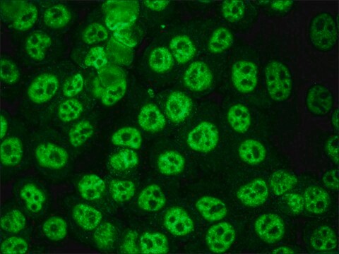
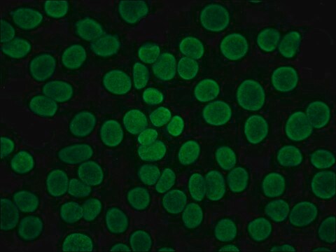
immunofluorescence: 1:64-1:128 using Hep2 cells
storage temp.
−20°C
target post-translational modification
unmodified
General description
IgG antibody plays a crucial role in humoral immune responses such as complement activation, phagocytosis, placental transport and cell surface-receptor binding. FITC (fluorescein isothiocyanate), a fluorochrome dye conjugation is useful in localization of antigen-antibody interaction of biological structures like cells or tissues. Anti-Human IgG (whole molecule) −FITC antibody diluted 1:100 in wash buffer is used in flow cytometry to analyze antibody affinity and virus binding. This antibody can also be used in indirect immunofluorescence antibody test. FITC labeled anti-human IgG antibody reacts specifically with human.
IgG antibody subtype is the most abundant serum immunoglobulins of the immune system. It is secreted by B cells and is found in blood and extracellular fluids and provides protection from infections caused by bacteria, fungi and viruses. Maternal IgG is transferred to fetus through the placenta that is vital for immune defence of the neonate against infections. Anti-Human IgG (whole molecule)−FITC antibody is specific for human IgG. Rabbit Anti-Human IgG is conjugated to crystalline fluorescein isothiocyanate (FITC) in an alkaline reaction and then further purified to remove free FITC.
Immunogen
IgG from pooled normal human serum
Application
Anti-Human IgG (whole molecule) −FITC antibody may be used as secondary antibody in indirect immunofluorescence and is also used in flow cytometry .
Applications in which this antibody has been used successfully, and the associated peer-reviewed papers, are given below.
Immunofluorescence (1 paper)
Immunofluorescence (1 paper)
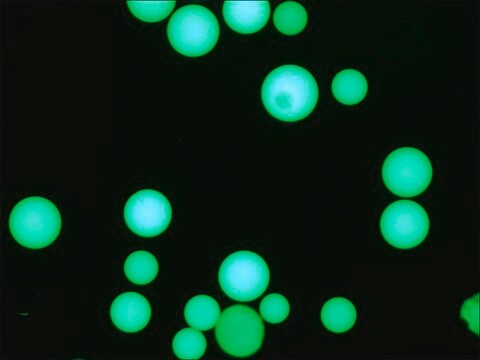
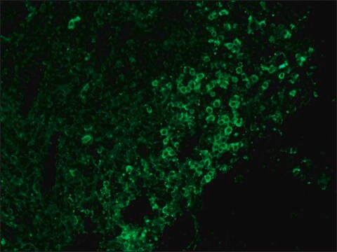
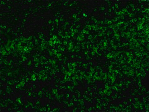
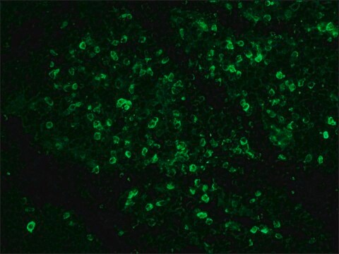
The antibody may be used for immunofluorescent staining of human peripheral blood lymphocytes at a working dilution of 1:64. A minimum working dilution of 1:64 was used on acetone fixed mouse liver cells and anti-nuclear antibody positive serum as the primary antibody. For flow cytometry to label NIH3T3 cells, the antibody was used at a working dilution of 1:100. The antibody was also used in ELISA using human sera.
Physical form
Solution in 0.01 M phosphate buffered saline, pH 7.4, containing 15 mM sodium azide.
Disclaimer
Unless otherwise stated in our catalog or other company documentation accompanying the product(s), our products are intended for research use only and are not to be used for any other purpose, which includes but is not limited to, unauthorized commercial uses, in vitro diagnostic uses, ex vivo or in vivo therapeutic uses or any type of consumption or application to humans or animals.
Not finding the right product?
Try our Product Selector Tool.
Storage Class Code
10 - Combustible liquids
WGK
nwg
Flash Point(F)
Not applicable
Flash Point(C)
Not applicable
Personal Protective Equipment
dust mask type N95 (US), Eyeshields, Gloves
Choose from one of the most recent versions:
Already Own This Product?
Find documentation for the products that you have recently purchased in the Document Library.
Customers Also Viewed
Bahador Sarkari et al.
Interdisciplinary perspectives on infectious diseases, 2014, 505134-505134 (2014-09-02)
Serological assays have been extensively evaluated for diagnosis of visceral leishmaniasis (VL) and considered as a routine method for diagnosis of VL while these methods are not properly evaluated for diagnosis of cutaneous leishmaniasis (CL). This study aimed to assess
Cherry C Chen et al.
Biomaterials, 32(27), 6579-6587 (2011-06-21)
Complement fixation to surface-conjugated ligands plays a critical role in determining the fate of targeted colloidal particles after intravenous injection. In the present study, we examined the immunogenicity of targeted microbubbles with various surface architectures and ligand surface densities using
Detection of Infectious Rotaviruses by Flow Cytometry .
Albert Bosch, Rosa M. Pinto
Springer Semin. Immunopath., 268(2), 61-68 (2004)
Evaluation of serological tests for the diagnosis of visceral Leishmaniasis
Kilic S et al
Turkish Journal of Medical Sciences, 38, 13-19 (2008)
Albert Bosch et al.
Methods in molecular biology (Clifton, N.J.), 268, 61-68 (2004-05-25)
Human rotaviruses are considered the main cause of viral gastroenteritis in infants and young children throughout the world. Their transmission is through the fecal-oral route, mostly after ingestion of contaminated water and food. Since an extremely high number of virus
Our team of scientists has experience in all areas of research including Life Science, Material Science, Chemical Synthesis, Chromatography, Analytical and many others.
Contact Technical Service
