Skip to Content
Merck
All Photos(2)

Key Documents

MAB361

Sigma-Aldrich

Anti-Tau Antibody, a.a. 210-241, clone Tau-5

ascites fluid, clone Tau-5, Chemicon®

Synonym(s):

G protein beta1/gamma2 subunit-interacting factor 1, Neurofibrillary tangle protein, Paired helical filament-tau, microtubule-associated protein tau, microtubule-associated protein tau, isoform 4

Sign Into View Organizational & Contract Pricing


About This Item

UNSPSC Code:
12352203
eCl@ss:
32160702
NACRES:
NA.41

biological source

mouse

Quality Level

antibody form

ascites fluid

antibody product type

primary antibodies

clone

Tau-5, monoclonal

species reactivity

mouse, human, rat, bovine

manufacturer/tradename

Chemicon®

technique(s)

immunohistochemistry: suitable
western blot: suitable

isotype

IgG1

NCBI accession no.

UniProt accession no.

shipped in

dry ice

target post-translational modification

unmodified

Gene Information

General description

Microtubule Associated Proteins, or MAPS, bind to the tubulin subunits of microtubule structures and regulate their functional stability. In the cell MAPs bind to monomer and multimerized tubulin. MAP binding to multimerized tubulin further stabilizes the formation of higher order microtubulin structures. MAP binding to microtubule structures is mediated through phosphorylation through Microtubule Affinity Regulated Kinase (MARK). Phosphorylation releases MAPs bound to microtubules, destabilizing the structure, driving it toward disassembly. There are predominately two MAP types, I, II. Type II MAP includes MAP2, MAP4, and tau and are found in nervous tissue. Six tau isoforms exist in brain tissue, and they are distinguished by their number of binding domains. Three isoforms have three binding domains and the other three have four binding domains. The binding domains are located in the carboxy-terminus of the protein and are positively-charged (allowing it to bind to the negatively-charged microtubule). The isoforms with four binding domains are better at stabilizing microtubules than those with three binding domains.

Specificity

Tau from bovine, rat and human.

Immunogen

Bovine tau
Epitope: a.a. 210-241

Application

Anti-Tau Antibody, a.a. 210-241, clone Tau-5 detects level of Tau & has been published & validated for use in IH & WB.
Immunohistochemistry:
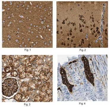
A previous lot of this antibody was used in immunohistochemistry.

Immunoblot:
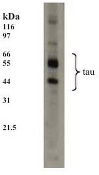
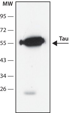
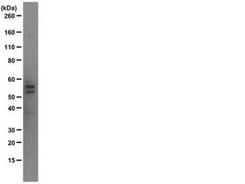
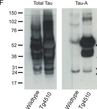
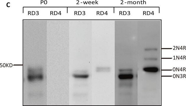
Recognizes phosphorylated and non-phosphorylated isoforms of tau ranging in size from 45-68 kDa.

Optimal working dilutions must be determined by end user.
Research Category
Neuroscience
Research Sub Category
Neurodegenerative Diseases

Quality

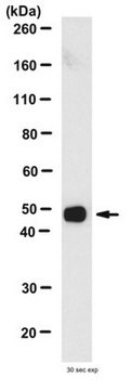
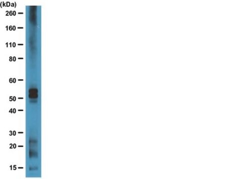
Routinely evaluated by Western Blot on PC12 lysates.

Western Blot Analysis:
1:500 dilution of this lot detected Tau on 10 μg of PC12 lysates.

Target description

45-68 kDa

Linkage

Replaces: MAB10417

Physical form

Unpurified
Unpurified mouse monoclonal IgG1 ascites fluid containing no preservatives.

Storage and Stability

Stable for 1 year at -20ºC in undiluted aliquots from date of receipt.
Handling Recommendations: Upon receipt, and prior to removing the cap, centrifuge the vial and gently mix the solution. Aliquot into microcentrifuge tubes and store at -20°C. Avoid repeated freeze/thaw cycles, which may damage IgG and affect product performance.

Analysis Note

Control
Brain Tissue, PC12 cell lysate

Other Notes

Concentration: Please refer to the Certificate of Analysis for the lot-specific concentration.

Legal Information

CHEMICON is a registered trademark of Merck KGaA, Darmstadt, Germany

Disclaimer

Unless otherwise stated in our catalog or other company documentation accompanying the product(s), our products are intended for research use only and are not to be used for any other purpose, which includes but is not limited to, unauthorized commercial uses, in vitro diagnostic uses, ex vivo or in vivo therapeutic uses or any type of consumption or application to humans or animals.

Not finding the right product?  

Try our Product Selector Tool.

Storage Class Code

10 - Combustible liquids

WGK

WGK 1

Flash Point(F)

Not applicable

Flash Point(C)

Not applicable


Certificates of Analysis (COA)

Search for Certificates of Analysis (COA) by entering the products Lot/Batch Number. Lot and Batch Numbers can be found on a product’s label following the words ‘Lot’ or ‘Batch’.

Already Own This Product?

Find documentation for the products that you have recently purchased in the Document Library.

Visit the Document Library

Mark D Johnson et al.
The Journal of biological chemistry, 279(25), 26685-26697 (2004-04-03)
Isotope-coded affinity tag reagents and high throughput mass spectrometry were used to quantitate changes in the expression of 150 proteins in mouse wild-type (p53(+/+)) cortical neurons undergoing DNA damage-induced death. Immunological techniques confirmed several of the changes in protein expression
Functional implications for the microtubule-associated protein tau: localization in oligodendrocytes.
LoPresti, P, et al.
Proceedings of the National Academy of Sciences of the USA, 92, 10369-10373 (1995)
Tau truncation during neurofibrillary tangle evolution in Alzheimer's disease.
Guillozet-Bongaarts, Angela L, et al.
Neurobiology of Aging, 26, 1015-1022 (2005)
Richard C Davis et al.
Acta neuropathologica, 115(2), 205-217 (2007-11-06)
Hirano bodies are paracrystalline F-actin-rich aggregations associated with a variety of conditions including aging, and neurodegenerative diseases. The composition and structure of these inclusions have been described by immunohistochemistry and ultrastructure, respectively. However, studies of the physiological function and dynamics
Elevated tauopathy and alpha-synuclein pathology in postmortem Parkinson's disease brains with and without dementia.
Wills, J; Jones, J; Haggerty, T; Duka, V; Joyce, JN; Sidhu, A
Experimental neurology null

Our team of scientists has experience in all areas of research including Life Science, Material Science, Chemical Synthesis, Chromatography, Analytical and many others.

Contact Technical Service