41325
Mayer′s Mucicarmine Stain Solution
for microscopy
Synonym(s):
Mucicarmine Stain Solution according to Mayer
Sign Into View Organizational & Contract Pricing
All Photos(1)
About This Item
Recommended Products
grade
for microscopy
Quality Level
product line
BioChemika
form
liquid
shelf life
limited shelf life, expiry date on the label
technique(s)
microbe id | staining: suitable
application(s)
food and beverages
hematology
histology
storage temp.
room temp
suitability
fungi
General description
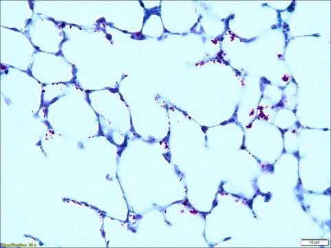
Most mucins contain serveral glycoproteins and can therefore be detected by stains for carbohydrates. Best carmine stain is specifically used for glycogens. Mucicarmine is an empirical stain. When applied for fungal staining, it stains the spores and hyphae as pale to dark red.
Components
Composition:
Carmine 1 g, Aluminum chloride 0.5 g, water 2 ml
Carmine 1 g, Aluminum chloride 0.5 g, water 2 ml
Not finding the right product?
Try our Product Selector Tool.
Signal Word
Danger
Hazard Statements
Precautionary Statements
Hazard Classifications
Eye Dam. 1 - Skin Corr. 1B
Supplementary Hazards
Storage Class Code
8B - Non-combustible corrosive hazardous materials
WGK
WGK 1
Flash Point(F)
Not applicable
Flash Point(C)
Not applicable
Personal Protective Equipment
dust mask type N95 (US), Eyeshields, Gloves
Choose from one of the most recent versions:
Already Own This Product?
Find documentation for the products that you have recently purchased in the Document Library.
Customers Also Viewed
O E Battles et al.
American journal of clinical pathology, 108(1), 6-12 (1997-07-01)
To identify a sensitive marker for extramammary Paget's disease and to identify histochemical and immunohistochemical features that suggest occult pelvic cancer in patients with extramammary Paget's disease, we retrieved all cases between 1983 and 1992 with a Standardized Nomenclature of
Alexandra Flávia Gazzoni et al.
Mycopathologia, 167(4), 197-202 (2008-12-05)
Here we report an unusual case of disseminated cryptococcosis in a patient with AIDS. Although typical Cryptococcus neoformans micromorphology was observed in tongue biopsy, cervical lymph node examination revealed atypical histopathologic findings. These included pseudohyphae, chains of budding yeasts and
Ji Kon Ryu et al.
Diagnostic cytopathology, 31(2), 100-105 (2004-07-30)
Pancreatic cystic neoplasms comprise a pathologically heterogeneous group with many shared clinical features. We assessed the reliability of cyst fluid analysis for the differential diagnosis of pancreatic cysts. Cyst fluid was obtained by fine-needle aspiration from 78 pancreatic cysts. The
Oluwole Fadare
Pathology international, 56(11), 683-687 (2006-10-17)
Some examples of lobular carcinoma in situ (LCIS) may be composed in part of signet ring cells. Such proliferations have been considered examples of pleomorphic LCIS based on pathological features of the more conventional component. However, the occurrence of LCIS
S P Hammar et al.
Ultrastructural pathology, 20(4), 293-325 (1996-07-01)
Pathologists routinely use histochemistry, immunohistochemistry, and electron microscopy to differentiate epithelial mesotheliomas from pulmonary adenocarcinomas. Epithelial mesotheliomas are usually mucicarmine-, PAS-diastase, and carcinoembryonic antigen-negative, whereas about 60-75% of pulmonary adenocarcinomas are mucicarmine- and PAS-diastase-positive, and about 90% express polyclonal carcinoembryonic
Our team of scientists has experience in all areas of research including Life Science, Material Science, Chemical Synthesis, Chromatography, Analytical and many others.
Contact Technical Service

