Skip to Content
Merck
All Photos(5)

Key Documents

HPA009975

Sigma-Aldrich

Anti-RMDN3 antibody produced in rabbit

Prestige Antibodies® Powered by Atlas Antibodies, affinity isolated antibody, buffered aqueous glycerol solution

Synonym(s):

Anti-FAM82A2, Anti-FAM82C antibody produced in rabbit, Anti-family with sequence similarity 82, member C antibody produced in rabbit

Sign Into View Organizational & Contract Pricing


About This Item

UNSPSC Code:
12352203
Human Protein Atlas Number:
NACRES:
NA.41

biological source

rabbit

Quality Level

conjugate

unconjugated

antibody form

affinity isolated antibody

antibody product type

primary antibodies

clone

polyclonal

product line

Prestige Antibodies® Powered by Atlas Antibodies

form

buffered aqueous glycerol solution

species reactivity

human

technique(s)

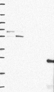
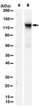
immunoblotting: 0.04-0.4 μg/mL
immunofluorescence: 0.25-2 μg/mL
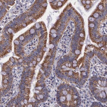
immunohistochemistry: 1:500-1:1000

immunogen sequence

DEVSCETVKMGRKDSLDLEEEAASGASSALEAGGSSGLEDVLPLLQQADELHRGDEQGKREGFQLLLNNKLVYGSRQDFLWRLARAYSDMCELTEEVSEKKSYALDGKEEAEAALEKGDESADCHLWYAVLCG

shipped in

wet ice

storage temp.

−20°C

target post-translational modification

unmodified

Gene Information

human ... FAM82A2(55177)

General description

RMDN3 (regulator of microtubule dynamics 3) was first identified by using a bait protein TCPTP (T-cell protein tyrosine phosphatase) in a yeast two hybrid screen. It has two binding sites for 14-3-3 proteins called conserved region 1 and conserved region 2. This protein has a molecular weight of 52,118Da, and is composed of 470 amino acids. This gene is localized to human chromosome 15q15.1, and is composed of 12 exons.

Immunogen

Protein FAM82C (Protein tyrosine phosphatase-interacting protein 51) (TCPTP-interacting protein 51) (Cerebral protein 10).

Application

All Prestige Antibodies Powered by Atlas Antibodies are developed and validated by the Human Protein Atlas (HPA) project and as a result, are supported by the most extensive characterization in the industry.

The Human Protein Atlas project can be subdivided into three efforts: Human Tissue Atlas, Cancer Atlas, and Human Cell Atlas. The antibodies that have been generated in support of the Tissue and Cancer Atlas projects have been tested by immunohistochemistry against hundreds of normal and disease tissues and through the recent efforts of the Human Cell Atlas project, many have been characterized by immunofluorescence to map the human proteome not only at the tissue level but now at the subcellular level. These images and the collection of this vast data set can be viewed on the Human Protein Atlas (HPA) site by clicking on the Image Gallery link. We also provide Prestige Antibodies® protocols and other useful information.

Biochem/physiol Actions

RMDN3 (regulator of microtubule dynamics 3) protein is involved in cell differentiation and apoptosis. Methylation status of this gene′s promoter determines its expression, which is increased in prostate cancer. In human muscle cells, the expression of this protein is dependent on the differentiation stage. The increase in the expression of this protein is correlated with the increase in proteins involved in advanced stages of myotubal differentiation, namely sarcoplasmic reticulum Ca2+ ATPase and myosin heavy-chain protein. RMDN3 is expressed in basal cell carcinoma, squamous cell carcinoma and the peritumoral tissue, and thus, might be a key player in the pathogenesis of keratinocyte carcinoma.

Features and Benefits

Prestige Antibodies® are highly characterized and extensively validated antibodies with the added benefit of all available characterization data for each target being accessible via the Human Protein Atlas portal linked just below the product name at the top of this page. The uniqueness and low cross-reactivity of the Prestige Antibodies® to other proteins are due to a thorough selection of antigen regions, affinity purification, and stringent selection. Prestige antigen controls are available for every corresponding Prestige Antibody and can be found in the linkage section.

Every Prestige Antibody is tested in the following ways:
  • IHC tissue array of 44 normal human tissues and 20 of the most common cancer type tissues.
  • Protein array of 364 human recombinant protein fragments.

Linkage

Corresponding Antigen APREST71998

Physical form

Solution in phosphate-buffered saline, pH 7.2, containing 40% glycerol and 0.02% sodium azide

Legal Information

Prestige Antibodies is a registered trademark of Merck KGaA, Darmstadt, Germany

Disclaimer

Unless otherwise stated in our catalog or other company documentation accompanying the product(s), our products are intended for research use only and are not to be used for any other purpose, which includes but is not limited to, unauthorized commercial uses, in vitro diagnostic uses, ex vivo or in vivo therapeutic uses or any type of consumption or application to humans or animals.

Not finding the right product?  

Try our Product Selector Tool.

Storage Class Code

10 - Combustible liquids

WGK

WGK 1

Flash Point(F)

Not applicable

Flash Point(C)

Not applicable

Personal Protective Equipment

dust mask type N95 (US), Eyeshields, Gloves

Choose from one of the most recent versions:

Certificates of Analysis (COA)

Lot/Batch Number

Don't see the Right Version?

If you require a particular version, you can look up a specific certificate by the Lot or Batch number.

Already Own This Product?

Find documentation for the products that you have recently purchased in the Document Library.

Visit the Document Library

P Koch et al.
Journal of cellular and molecular medicine, 12(5B), 2083-2095 (2008-11-18)
The novel protein PTPIP51 (SwissProt accession code Q96SD6) is known to interact with two non-transmembrane protein-tyrosine phosphatases, PTP1B and TCPTP in vitro. Overexpression of the full-length protein induces apoptosis in HeLa and HEK293T cells (Lv et al. 2006). PTPIP51 shows
Philipp Koch et al.
The Prostate, 69(16), 1751-1762 (2009-08-20)
Protein tyrosine phosphatase interacting protein 51 (PTPIP51) shows a tissue-specific expression pattern and is associated with cellular differentiation and apoptosis in several mammalian tissues. Overexpression of the full-length protein enhances apoptosis. It is also expressed in various carcinomas. In this
Justus Barop et al.
The journal of histochemistry and cytochemistry : official journal of the Histochemistry Society, 57(5), 425-435 (2009-01-07)
Protein tyrosine phosphatase-interacting protein 51 (PTPIP51) expression was analyzed in proliferating and differentiating human myogenic cells cultured in vitro. Satellite cell cultures derived from four different individuals were used in this study. To analyze the expression of PTPIP51, myoblasts were
Laurence Walch et al.
Scientific reports, 8(1), 17121-17121 (2018-11-22)
The spatial organization of cells depends on coordination between cytoskeletal systems and intracellular organelles. The Arf1 small G protein and its activator GBF1 are important regulators of Golgi organization, maintaining its morphology and function. Here we show that GBF1 and
Kurt J De Vos et al.
Human molecular genetics, 21(6), 1299-1311 (2011-12-02)
A proline to serine substitution at position 56 in the gene encoding vesicle-associated membrane protein-associated protein B (VAPB) causes some dominantly inherited familial forms of motor neuron disease including amyotrophic lateral sclerosis (ALS) type-8. VAPB is an integral endoplasmic reticulum

Our team of scientists has experience in all areas of research including Life Science, Material Science, Chemical Synthesis, Chromatography, Analytical and many others.

Contact Technical Service