Skip to Content
Merck
All Photos(1)

Key Documents

120R-1

Sigma-Aldrich

CD20 (SP32) Rabbit Monoclonal Antibody

Sign Into View Organizational & Contract Pricing


About This Item

UNSPSC Code:
12352203
NACRES:
NA.41
clone:
SP32, monoclonal
application:
IHC (p)
species reactivity:
human
technique(s):
immunohistochemistry (formalin-fixed, paraffin-embedded sections): 1:100-1:500
citations:
5

biological source

rabbit

Quality Level

100
500

conjugate

unconjugated

antibody form

culture supernatant

antibody product type

primary antibodies

clone

SP32, monoclonal

description

For In Vitro Diagnostic Use in Select Regions (See Chart)

form

buffered aqueous solution

species reactivity

human

packaging

vial of 0.1 mL concentrate (120R-14)
vial of 0.5 mL concentrate (120R-15)
bottle of 1.0 mL predilute (120R-17)
vial of 1.0 mL concentrate (120R-16)
bottle of 7.0 mL predilute (120R-18)

manufacturer/tradename

Cell Marque

technique(s)

immunohistochemistry (formalin-fixed, paraffin-embedded sections): 1:100-1:500

isotype

IgG1

control

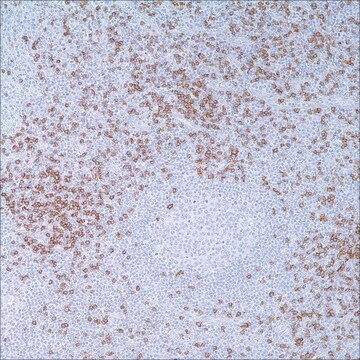
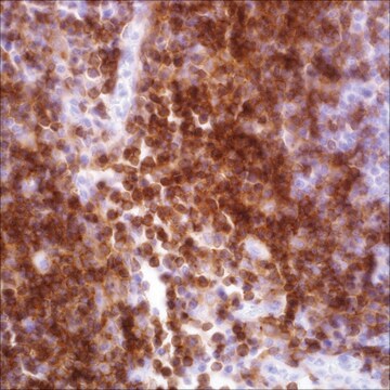
tonsil

shipped in

wet ice

storage temp.

2-8°C

visualization

membranous

General description

Anti-CD20 (pan B-cell) reacts with a membrane antigen that is present in B-cells as this epitope is acquired late in the pre-B-cell stage of maturation and remains on cells throughout most of the differentiation process, although it is lost at the plasma cell stage. This antibody recognizes Reed-Sternberg cells, predominant in classic Hodgkin′s disease, in approximately 20% of cases, and approximately half of lymphoblastic lymphoma/leukemias. Since no staining of histiocytes or plasma cells has been observed and CD20 has rarely been detected in T-cell malignancies, it is a dependable marker of B-cell lymphomas. Anti-CD20 does not cross-react with non-hematopoetic neoplasms.

Linkage

CD20 Positive Control Slides, Product No. 120S, are available for immunohistochemistry (formalin-fixed, paraffin-embedded sections).

Physical form

Solution in Tris Buffer, pH 7.3-7.7, with 1% BSA and <0.1% Sodium Azide

Preparation Note

Download the IFU specific to your product lot and formatNote: This requires a keycode which can be found on your packaging or product label.

Other Notes

For Technical Service please contact: 800-665-7284 or email: service@cellmarque.com

Legal Information

Cell Marque is a trademark of Merck KGaA, Darmstadt, Germany

Not finding the right product?  

Try our Product Selector Tool.

Storage Class Code

12 - Non Combustible Liquids

WGK

WGK 2

Flash Point(F)

Not applicable

Flash Point(C)

Not applicable


Choose from one of the most recent versions:

Certificates of Analysis (COA)

Lot/Batch Number

Don't see the Right Version?

If you require a particular version, you can look up a specific certificate by the Lot or Batch number.

Already Own This Product?

Find documentation for the products that you have recently purchased in the Document Library.

Visit the Document Library

P Korkolopoulou et al.
Histopathology, 24(6), 511-515 (1994-06-01)
Recent evidence indicates that membrane-bound immunoglobulin on B lymphocytes is associated with a molecule which comprises the products of the mb-1 and B29 genes. This molecule is a highly specific marker for B-cells, presumably because of its central functional role
G S Pinkus et al.
The American journal of pathology, 133(2), 211-217 (1988-11-01)
Immunoreactivity for L26, a highly effective pan B cell marker that can be detected in paraffin sections, was evaluated in 72 cases of Hodgkin's disease of various histologic types. In all cases of nodular lymphocyte predominance type of Hodgkin's disease
Diagnostic Immunohistochemistry: Theranostic and Genomic Applications (2006)
A L Epstein et al.
Journal of immunology (Baltimore, Md. : 1950), 133(2), 1028-1036 (1984-08-01)
Two monoclonal antibodies (LN-1, LN-2) reactive with B lymphocytes in B5 formalin-fixed, paraffin-embedded tissue sections have been produced by utilizing cell extracts from pokeweed mitogen-stimulated peripheral blood lymphocytes and diffuse histiocytic lymphoma SU-DHL-4 cells, respectively. Both monoclonal antibodies were initially
R J Marder et al.
Laboratory investigation; a journal of technical methods and pathology, 52(5), 497-504 (1985-05-01)
Monoclonal antibodies (MoAbs) to B cell- and T cell-specific antigens uniformly have been restricted in their use to cell suspension or frozen section techniques because the antigens that they identify are either masked or lost in the fixation or paraffin-embedding

Our team of scientists has experience in all areas of research including Life Science, Material Science, Chemical Synthesis, Chromatography, Analytical and many others.

Contact Technical Service