Skip to Content
Merck
All Photos(2)

Key Documents

T6674

Sigma-Aldrich

Tumor Necrosis Factor-α human

TNF-α, recombinant, expressed in E. coli, powder, suitable for cell culture

Synonym(s):

hTNF-α, TNF-α

Sign Into View Organizational & Contract Pricing


About This Item

CAS Number:
MDL number:
UNSPSC Code:
12352202
NACRES:
NA.77

biological source

human

Quality Level

recombinant

expressed in E. coli

Assay

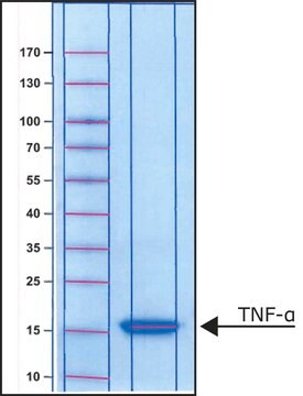
≥97% (SDS-PAGE)

form

powder

potency

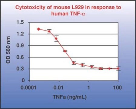
0.02-0.3 ng/mL ED50/EC50

quality

endotoxin tested

mol wt

~17.4 kDa

packaging

pkg of 5x10 μg
pkg of 10 μg

storage condition

avoid repeated freeze/thaw cycles

technique(s)

cell culture | mammalian: suitable

impurities

<1 EU/μg

UniProt accession no.

storage temp.

−20°C

Gene Information

human ... TNF(7124)

Looking for similar products? Visit Product Comparison Guide

Application

TNF-α include stimulating growth of human fibroblasts and other cell lines activating polymorphonuclear neutrophils and osteoclasts and inducing interleukin-1, prostaglandin E2, and collagenase production.
Tumor Necrosis Factor-α (TNFα) human has been used:
  • to analyze the effects of cytokine TNFα-stressed human neuronal and glial (HNG) cells
  • to investigate the molecular mechanisms of TNFα-mediated prolyl-4 hydroxylase α1 (P4Hα1) suppression
  • to induce death-receptor-mediated apoptosis in HeLa cells.

Biochem/physiol Actions

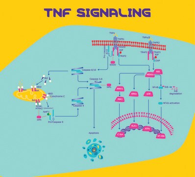
Tumore necrosis factor-α (TNF-α), also known as cachectin, is expressed as a 26 kDa membrane bound protein and is then cleaved by TNF-α converting enzyme (TACE) to release the soluble 17 kDa monomer, which forms homotrimers in circulation. TNF-α plays roles in antitumor activity, immune modulation, inflammation, anorexia, cachexia, septic shock, viral replication and hematopoiesis. TNF-α is expressed by a great variety of cells, with numerous inductive and suppressive agents. Primarily, TNF-α is produced by macrophages in response to immunological challenges such as bacteria (lipopolysaccharides), viruses, parasites, mitogens and other cytokines. TNF-α is cytotoxic for many transformed cells (its namesake activity) but in normal diploid cells, it can stimulate proliferation (fibroblasts), differentiation (myeloid cells) or activation (neutrophils). TNF-α also shows antiviral effects against both DNA and RNA viruses and it induces production of several other cytokines. Although TNF-α is used in clinical trials as an antitumor agent, Sigma′s cytokine, growth factor and hormone products are for research only. TNF-α and the related molecule TNF-β (LT-α) share close structural homology with 28% amino acid sequence identity and both activate the same TNF receptors, TNFR1 and TNFR2. Mouse and human TNF-α share 79% amino acid sequence identity. Unlike human TNF-α, the mouse form is N-glycosylated.

Physical form

Lyophilized from a 0.2 μm-filtered buffer solution containing 3mM Tris-HCl and 5% trehalose.

Analysis Note

The biological activity of TNF-α is measured by the cytolysis of murine L929 cells.

Certificates of Analysis (COA)

Search for Certificates of Analysis (COA) by entering the products Lot/Batch Number. Lot and Batch Numbers can be found on a product’s label following the words ‘Lot’ or ‘Batch’.

Already Own This Product?

Find documentation for the products that you have recently purchased in the Document Library.

Visit the Document Library

A cytomegalovirus-encoded inhibitor of apoptosis that suppresses caspase-8 activation
Skaletskaya A, et al.
Proceedings of the National Academy of Sciences of the USA, 98(14), 7829-7834 (2001)
miRNA-155 up-regulation and complement factor H (CFH) deficits in Down?s Syndrome
Li YY, et al.
Neuroreport, 23(3), 168-168 (2012)
Role of NonO-histone interaction in TNFalpha-suppressed Prolyl-4-hydroxylase alpha1
Zhang C, et al.
Biochimica et Biophysica Acta - Molecular Cell Research, 1783(8), 1517-1528 (2008)
Casper Steenholdt et al.
The American journal of gastroenterology, 109(7), 1055-1064 (2014-05-07)
Cost-effective guidance of therapeutic strategy in Crohn's disease patients with secondary infliximab (IFX) treatment failure may be achieved by serum IFX and anti-IFX antibody (Ab) measurements by radioimmunoassay (RIA). This study investigated implications of using other techniques for this purpose.
David J Konieczkowski et al.
Cancer discovery, 4(7), 816-827 (2014-04-29)
Most melanomas harbor oncogenic BRAF(V600) mutations, which constitutively activate the MAPK pathway. Although MAPK pathway inhibitors show clinical benefit in BRAF(V600)-mutant melanoma, it remains incompletely understood why 10% to 20% of patients fail to respond. Here, we show that RAF

Protocols

WST-1 assay protocol for measuring cell viability, proliferation, activation and cytotoxicity. Instructions for WST-1 reagent preparation and examples of applications. Frequently asked questions and troubleshooting guide for WST-1 assay.

Protocol Guide: XTT Assay for Cell Viability and Proliferation

Our team of scientists has experience in all areas of research including Life Science, Material Science, Chemical Synthesis, Chromatography, Analytical and many others.

Contact Technical Service