MCYTOMAG-70K
MILLIPLEX® Mouse Cytokine/Chemokine Magnetic Bead Panel - Immunology Multiplex Assay
Simultaneously analyze multiple cytokine and chemokine biomarkers with Bead-Based Multiplex Assays using the Luminex technology, in mouse serum, plasma and cell culture samples.
Synonym(s):
Luminex® Mouse cytokine immunoassay, Millipore Mouse cytokine immunoassay, Mouse cytokine Multiplex Assay
About This Item
Recommended Products
Quality Level
species reactivity
mouse
manufacturer/tradename
Milliplex®
assay range
accuracy: 85-107%
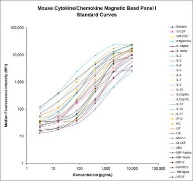
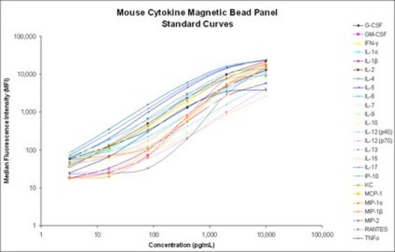
standard curve range: 3.2-10,000 pg/mL
technique(s)
multiplexing: suitable
detection method
fluorometric (Luminex xMAP)
shipped in
wet ice
General description
- More flexible plate and plate washer options
- Improved performance with turbid serum/plasma samples
- Assay results equivalent to non- beads
- Automated washing avoids many problems associated with vacuum filtration washing
MILLIPLEX® Mouse Cytokine / Chemokine panel enables you to focus on the therapeutic potential of cytokines as well as the modulation of cytokine expression. Coupled with the Luminex® xMAP® platform in a bead format, you receive the advantage of ideal speed and sensitivity, allowing quantitative multiplex detection of dozens of analytes simultaneously, which can dramatically improve productivity.
Panel Type: Cytokines/Chemokines
Application
- Analytes: G-CSF, GM-CSF, IFN-γ, IL-1α, IL-1β, IL-2, IL-3, IL-4, IL-5, IL-6, IL-7, IL-9, IL-10, IL-12 (p40), IL-12 (p70), IL-13, IL-15, IL-17, IP-10, KC, LIF, LIX, MCP-1, M-CSF, MIG, MIP-1α, MIP-1β, MIP-2, RANTES, TNF-α, VEGF, Eotaxin/CCL11
Features and Benefits
Other Notes
Legal Information
Signal Word
Danger
Hazard Statements
Precautionary Statements
Hazard Classifications
Acute Tox. 4 Dermal - Acute Tox. 4 Inhalation - Acute Tox. 4 Oral - Aquatic Chronic 2 - Eye Dam. 1 - Skin Sens. 1 - STOT RE 2
Target Organs
Respiratory Tract
Storage Class Code
10 - Combustible liquids
Certificates of Analysis (COA)
Search for Certificates of Analysis (COA) by entering the products Lot/Batch Number. Lot and Batch Numbers can be found on a product’s label following the words ‘Lot’ or ‘Batch’.
Already Own This Product?
Find documentation for the products that you have recently purchased in the Document Library.
Customers Also Viewed
Related Content
Learn how immunoassays are used in cosmetics and personal care research to discover potential harmful effects of cosmetics products, and assess markers of inflammation, sensitization, aging, and tissue regeneration among others.
See how multiplexing the inflammation signaling pathway with MILLIPLEX® inflammation assays or cell signaling assays can help researchers bridge the gap between immunology and cell signaling, including investigating T cell signaling, Th Cell differentiation, inflammatory response signaling, and sepsis signaling.
Our team of scientists has experience in all areas of research including Life Science, Material Science, Chemical Synthesis, Chromatography, Analytical and many others.
Contact Technical Service
