Skip to Content
Merck
All Photos(1)

Documents

ABS1646

Sigma-Aldrich

Anti-VPS4A (UT289)

serum, from rabbit

Synonym(s):

Vacuolar protein sorting-associated protein 4A, Protein SKD2, SKD1-homolog, Vacuolar protein sorting factor 4A, Vacuolar sorting protein 4, VPS4A

Sign Into View Organizational & Contract Pricing


About This Item

UNSPSC Code:
12352203
eCl@ss:
32160702

biological source

rabbit

Quality Level

antibody form

serum

antibody product type

primary antibodies

clone

polyclonal

species reactivity

human, mouse

technique(s)

immunocytochemistry: suitable
immunoprecipitation (IP): suitable
western blot: suitable

NCBI accession no.

UniProt accession no.

shipped in

ambient

target post-translational modification

unmodified

Gene Information

human ... VPS4A(27183)
mouse ... Vps4A(116733)

General description

Vacuolar protein sorting-associated protein 4A (EC 3.6.4.6; UniProt Q9UN37; also known as hVPS4, Protein SKD2, SKD1-homolog, Vacuolar protein sorting factor 4A, Vacuolar sorting protein 4, VPS4A) is encoded by the VPS4A (also known as SKD1A, VPS4, VPS4-1) gene (Gene ID 27183) in human. VPS4A is a member of the ATPases associated with various cellular activities (AAA) family proteins. The ESCRT (endosomal sorting complexes required for transport) machinery consists of five complexes (SCRT-0, ESCRT-I, ESCRT-II, ESCRT-III, and Vps4) and mediates multiple cellular membrane remodeling events, ranging from multivesicular bodies and exosomes formation to the final membrane abscission stage of cytokinesis, all of which converge in the scission of the narrow membrane neck that connects bud to parent membrane or daughter cell to daughter cell. Scission depends on the polymerization of soluble ESCRT-III monomers or dimers into tightly membrane-bound filaments. VPS4A is essential for the functions of the ESCRTs in cells by mediating the disassembly of ESCRT-III filaments to ensure a constant replenish of the pool of soluble ESCRT-III monomers. The ESCRTs are also exploited by HIV-1 and many other enveloped viruses for their escape from the host cells. In addition, VPS4A is reported to facilitate the secretion of oncogenic miRNAs in exosomes and downregulation of VPS4A in hepatocellular carcinoma (HCC) is associated with tumor progression and metastasis.

Specificity

VPS4A immunostaining and target band detection by UT289 was observed only in untransfected, but not VPS4A shRNA-transfected HeLa cells (Morita, E., et al. (2010). Proc. Natl. Acad. Sci. U.S.A. 107(29):12889-12894). Due to a typographical error, UT289 was listed as UT829 in Table S1 of Skalicky, J.J., et al. (2012). J. Biol. Chem. 287(52):43910-43926.

Immunogen

Recombinant human VPS4A lacking N-terminal MIT domain (Morita, E., et al. (2010). Proc. Natl. Acad. Sci. U.S.A. 107(29):12889-12894).

Application

Detect Vacuolar protein sorting-associated protein 4A using this rabbit polyclonal Anti-VPS4A (UT289), Cat. No. ABS1646, validated for use in Immunocytochemistry, Immunoprecipitation, and Western Blotting.
Immunocytochemistry Analysis: A representative lot detected VPS4A immunoreactivity concentrated at the spindle pole during metaphase and at the midbody during cytokinesis in 3% paraformaldehyde-fixed HeLa cells. No staining was seen in VPS4A shRNA-transfected HeLa cells (Morita, E., et al. (2010). Proc. Natl. Acad. Sci. U.S.A. 107(29):12889-12894).

Immunoprecipitation Analysis: A representative lot immunoprecipited EGFP-VPS4A fusion together with CHMP5-Myc and Myc-LIP5 exogenously expressed in HEK293T cells. CHMP5-Myc with L4D mutation and Myc-LIP5 with M64A, W147D, or Y278A mutation failed to co-immunoprecipitate with VPS4A (Skalicky, J.J., et al. (2012). J. Biol. Chem. 287(52):43910-43926).

Western Blotting Analysis: A representative lot detected both endogenous VPS4A and exogenously expressed EGFP-VPS4A fusion in HEK293T cell lysates as well as in Anti-Myc immunoprecipites from CHMP5-Myc or Myc-LIP5 expressing cells (Skalicky, J.J., et al. (2012). J. Biol. Chem. 287(52):43910-43926).

Western Blotting Analysis: A representative lot detected the VPS4A target band in lysate from untransfected, but not VPS4A shRNA-transfected, HeLa cells (Morita, E., et al. (2010). Proc. Natl. Acad. Sci. U.S.A. 107(29):12889-12894).
Research Category
Signaling

Quality

Evaluated by Western Blotting in MCF-7 cell lysate.

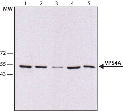
Western Blotting Analysis: A 1:1,000 dilution of this antiserum detected VPS4A in 10 µg of MCF-7 cell lysate.

Target description

~52 kDa observed. 48.90/48.91 kDa (human/mouse) calculated. Uncharacterized bands may be observed in some lysate(s).

Physical form

Rabbit polyclonal antibody serum with 0.05% sodium azide.
Unpurified.

Storage and Stability

Stable for 1 year at -20°C from date of receipt.
Handling Recommendations: Upon receipt and prior to removing the cap, centrifuge the vial and gently mix the solution. Aliquot into microcentrifuge tubes and store at -20°C. Avoid repeated freeze/thaw cycles, which may damage IgG and affect product performance.

Other Notes

Concentration: Please refer to lot specific datasheet.

Disclaimer

Unless otherwise stated in our catalog or other company documentation accompanying the product(s), our products are intended for research use only and are not to be used for any other purpose, which includes but is not limited to, unauthorized commercial uses, in vitro diagnostic uses, ex vivo or in vivo therapeutic uses or any type of consumption or application to humans or animals.

Not finding the right product?  

Try our Product Selector Tool.

Storage Class Code

12 - Non Combustible Liquids

WGK

WGK 1


Certificates of Analysis (COA)

Search for Certificates of Analysis (COA) by entering the products Lot/Batch Number. Lot and Batch Numbers can be found on a product’s label following the words ‘Lot’ or ‘Batch’.

Already Own This Product?

Find documentation for the products that you have recently purchased in the Document Library.

Visit the Document Library

Lin Deng et al.
Journal of virology, 96(6), e0181121-e0181121 (2022-01-20)
We previously reported that hepatitis C virus (HCV) infection activates the reactive oxygen species (ROS)/c-Jun N-terminal kinase (JNK) signaling pathway. However, the roles of ROS/JNK activation in the HCV life cycle remain unclear. We sought to identify a novel role

Our team of scientists has experience in all areas of research including Life Science, Material Science, Chemical Synthesis, Chromatography, Analytical and many others.

Contact Technical Service