Skip to Content
Merck
All Photos(1)

Key Documents

04-772

Sigma-Aldrich

Anti-Src Antibody, CT, clone NL19 | 04-772

clone NL19, Upstate®, from rabbit

Synonym(s):

Proto-oncogene tyrosine-protein kinase SRC, Protooncogene SRC, Rous sarcoma tyrosine kinase pp60c-src, Tyrosine-protein kinase SRC-1, v-src sarcoma (Schmidt-Ruppin A-2) viral oncogene homolog (avian)

Sign Into View Organizational & Contract Pricing


About This Item

UNSPSC Code:
12352203
eCl@ss:
32160702
NACRES:
NA.41

biological source

rabbit

Quality Level

antibody form

culture supernatant

antibody product type

primary antibodies

clone

NL19, monoclonal

species reactivity

mouse, human, rat

manufacturer/tradename

Upstate®

technique(s)

immunoprecipitation (IP): suitable
western blot: suitable

isotype

IgG

NCBI accession no.

UniProt accession no.

shipped in

dry ice

target post-translational modification

unmodified

Gene Information

human ... SRC(6714)

General description

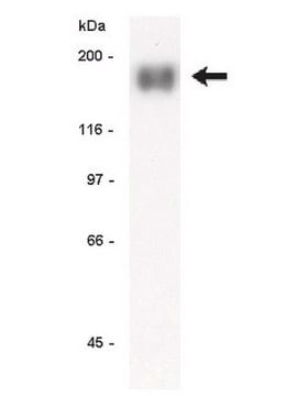
SRC-1 is a member of a gene family that functions in the full transcriptional activation of the steroid hormone receptor superfamily and contains intrinsic histone acetyltransferase activity. SRC-1 has a molecular weight of 160 kDa and has a basic helix-loop-helix (bHLH)-PAS domain.

Specificity

Predicted to cross react with frog and chicken. The immunogen sequence shares 22/23 amino acids with Yes and Fyn, and 21/23 amino acids with Lyn and Hck.
Src-tyrosine kinase family

Immunogen

KLH conjugated synthetic peptide (CKVADFGLARLIEDNEYTARQGA) of Src (avian sarcoma virus), residues 400-422. The immunogen sequence shares 22/23 amino acids with Yes and Fyn, and 21/23 amino acids with Lyn and Hck.

Application

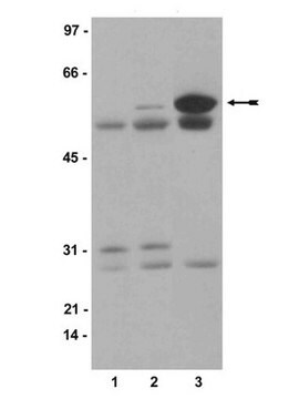
Western Blot Analysis: A 1:2000 to 1:8000 dilution of this lot detected Src in RIPA whole cell lysates from human A431 cells. Immunoprecipitation: 5-20 μL of a previous lot immunoprecipitated Src from 500 μg of A431 RIPA lysate.

Quality

routinely evaluated by immunoblot on RIPA whole cell lysates from human A431 and Jurkat cells, mouse 3T3/A31 cells or rat PC-12 cells

Target description

~53-62kDa

Linkage

Replaces: 05-772

Physical form

Format: Purified

Analysis Note

Control
RIPA whole cell lysates from human A431, Jurkat cells, mouse 3T3/A31 cells or rat PC-12 cells Included Positive Antigen Control: Catalog # 12-301, Non-Stimulated A431 cell lysate. Add 2.5 μL of 2-mercaptoethanol/100 μL of lysate and boil for 5 minutes to reduce the preparation. Load 20 μg of reduced lysate per lane for minigels.

Legal Information

UPSTATE is a registered trademark of Merck KGaA, Darmstadt, Germany

Not finding the right product?  

Try our Product Selector Tool.

Storage Class Code

12 - Non Combustible Liquids

WGK

WGK 1

Flash Point(F)

Not applicable

Flash Point(C)

Not applicable


Certificates of Analysis (COA)

Search for Certificates of Analysis (COA) by entering the products Lot/Batch Number. Lot and Batch Numbers can be found on a product’s label following the words ‘Lot’ or ‘Batch’.

Already Own This Product?

Find documentation for the products that you have recently purchased in the Document Library.

Visit the Document Library

Machika Kawamura et al.
Life science alliance, 4(8) (2021-06-26)
The pericentromeric heterochromatin of one-cell embryos forms a unique, ring-like structure around the nucleolar precursor body, which is absent in somatic cells. Here, we found that the histone H3 variants H3.1 and/or H3.2 (H3.1/H3.2) were localized asymmetrically between the male
Pazit Y Cohen et al.
American journal of respiratory cell and molecular biology, 40(2), 231-238 (2008-08-05)
We have previously demonstrated that myofibroblasts from lungs with bleomycin-induced fibrosis overexpress FasL molecules. Two subpopulations of fibroblasts, distinguished by their expression of Thy1 molecules, have been shown in the lungs of both mice and humans. Thy1-mediated FasL induction has
Amélie Juin et al.
The Journal of cell biology, 207(4), 517-533 (2014-11-26)
Accumulation of type I collagen fibrils in tumors is associated with an increased risk of metastasis. Invadosomes are F-actin structures able to degrade the extracellular matrix. We previously found that collagen I fibrils induced the formation of peculiar linear invadosomes

Our team of scientists has experience in all areas of research including Life Science, Material Science, Chemical Synthesis, Chromatography, Analytical and many others.

Contact Technical Service