Accéder au contenu
Merck
Toutes les photos(6)

Key Documents

HPA005466

Sigma-Aldrich

Anti-MYBBP1A antibody produced in rabbit

enhanced validation

Prestige Antibodies® Powered by Atlas Antibodies, affinity isolated antibody, buffered aqueous glycerol solution

Synonyme(s) :

Anti-FLJ37886, Anti-MYB binding protein (P160) 1a, Anti-P160, Anti-PAP2

Se connecterpour consulter vos tarifs contractuels et ceux de votre entreprise/organisme


About This Item

Code UNSPSC :
12352203
Numéro HPA (Human Protein Atlas):
Nomenclature NACRES :
NA.41

Source biologique

rabbit

Conjugué

unconjugated

Forme d'anticorps

affinity isolated antibody

Type de produit anticorps

primary antibodies

Clone

polyclonal

Gamme de produits

Prestige Antibodies® Powered by Atlas Antibodies

Forme

buffered aqueous glycerol solution

Espèces réactives

human

Validation améliorée

orthogonal RNAseq
Learn more about Antibody Enhanced Validation

Technique(s)

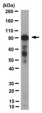
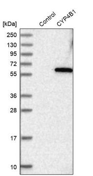
immunoblotting: 0.04-0.4 μg/mL
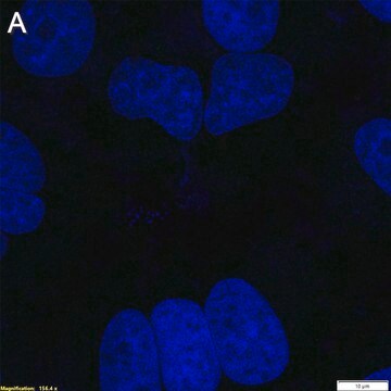
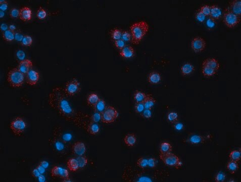
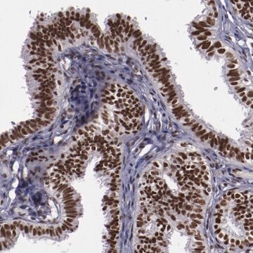
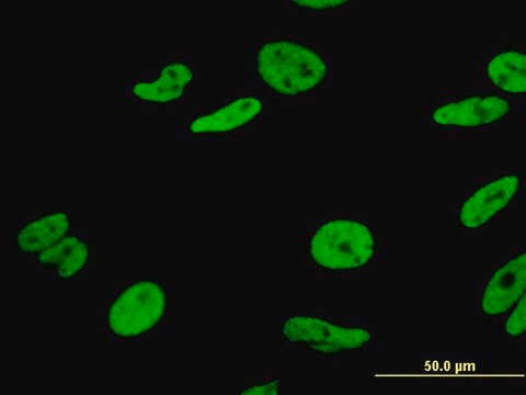
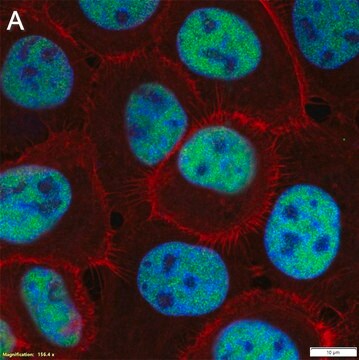
immunofluorescence: 0.25-2 μg/mL
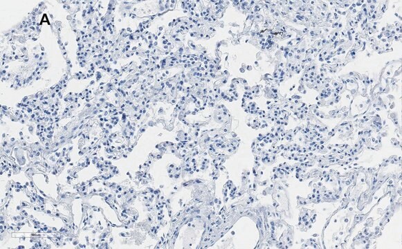
immunohistochemistry: 1:50-1:200

Séquence immunogène

EDLPLCSILQQIQEKYDLHQVKKAMLRPALFANLFGVLALFQSGRLVKDQEALMKSVKLLQALAQYQNHLQEQPRKALVDILSEVSKATLQEILPEVLKADLNIILSSPEQLELFLLAQQKVPSKLKKLVG

Numéro d'accès UniProt

Conditions d'expédition

wet ice

Température de stockage

−20°C

Modification post-traductionnelle de la cible

unmodified

Informations sur le gène

human ... MYBBP1A(10514)

Description générale

Myb-binding protein 1a (MYBBP1A) is a transcriptional co-regulator, which was originally identified as the protein which interacts with the proto-oncogene c-Myb. It is largely localized to the nucleolus, as it contains basic amino-acid repeats at its C-terminal. This protein weighs 160kDa and is expressed ubiquitously. Processing of the C-terminal nucleolar signal peptide produces two N-terminal forms of 140kDa and 67kDa. These forms are found in the nucleoplasm. MYBBP1A is located in the chromosomal region 17p13.3.

Immunogène

MYB binding protein (P160) 1a recombinant protein epitope signature tag (PrEST)

Application

All Prestige Antibodies Powered by Atlas Antibodies are developed and validated by the Human Protein Atlas (HPA) project and as a result, are supported by the most extensive characterization in the industry.

The Human Protein Atlas project can be subdivided into three efforts: Human Tissue Atlas, Cancer Atlas, and Human Cell Atlas. The antibodies that have been generated in support of the Tissue and Cancer Atlas projects have been tested by immunohistochemistry against hundreds of normal and disease tissues and through the recent efforts of the Human Cell Atlas project, many have been characterized by immunofluorescence to map the human proteome not only at the tissue level but now at the subcellular level. These images and the collection of this vast data set can be viewed on the Human Protein Atlas (HPA) site by clicking on the Image Gallery link. We also provide Prestige Antibodies® protocols and other useful information.

Actions biochimiques/physiologiques

Myb-binding protein 1a (MYBBP1A) regulates essential functions such as cell division, proliferation and apoptosis. It binds to NFκB via its RelA/p65 subunit, and prevents it trans-activating capacity. It binds to multiple transcription factors, such as PPARγ co-activator 1α, and inhibits them. This protein also forms a part of the Ret-CoR co-repressor complex. It activates the aryl hydrocarbon receptor (AhR). It induces apoptosis by preventing Mdm2 mediated degradation of p53. MYBBP1A maintains the silent state of rDNA and negatively regulates the epigenetic state of silent rDNA. It also negatively regulates the expression of rRNA. MYBBP1A prevents breast cancer tumorigenesis and controls the metastasis as well as proliferation of head and neck squamous cell carcinoma cells.

Caractéristiques et avantages

Prestige Antibodies® are highly characterized and extensively validated antibodies with the added benefit of all available characterization data for each target being accessible via the Human Protein Atlas portal linked just below the product name at the top of this page. The uniqueness and low cross-reactivity of the Prestige Antibodies® to other proteins are due to a thorough selection of antigen regions, affinity purification, and stringent selection. Prestige antigen controls are available for every corresponding Prestige Antibody and can be found in the linkage section.

Every Prestige Antibody is tested in the following ways:
  • IHC tissue array of 44 normal human tissues and 20 of the most common cancer type tissues.
  • Protein array of 364 human recombinant protein fragments.

Liaison

Corresponding Antigen APREST86952

Forme physique

Solution in phosphate buffered saline, pH 7.2, containing 40% glycerol and 0.02% sodium azide.

Informations légales

Prestige Antibodies is a registered trademark of Merck KGaA, Darmstadt, Germany

Clause de non-responsabilité

Unless otherwise stated in our catalog or other company documentation accompanying the product(s), our products are intended for research use only and are not to be used for any other purpose, which includes but is not limited to, unauthorized commercial uses, in vitro diagnostic uses, ex vivo or in vivo therapeutic uses or any type of consumption or application to humans or animals.

Vous ne trouvez pas le bon produit ?  

Essayez notre Outil de sélection de produits.

Code de la classe de stockage

10 - Combustible liquids

Classe de danger pour l'eau (WGK)

WGK 1

Point d'éclair (°F)

Not applicable

Point d'éclair (°C)

Not applicable


Certificats d'analyse (COA)

Recherchez un Certificats d'analyse (COA) en saisissant le numéro de lot du produit. Les numéros de lot figurent sur l'étiquette du produit après les mots "Lot" ou "Batch".

Déjà en possession de ce produit ?

Retrouvez la documentation relative aux produits que vous avez récemment achetés dans la Bibliothèque de documents.

Consulter la Bibliothèque de documents

Tetsuro Komatsu et al.
Traffic (Copenhagen, Denmark), 17(11), 1168-1180 (2016-10-27)
In adenoviral virions, the genome is organized into a chromatin-like structure by viral basic core proteins. Consequently viral DNAs must be replicated, chromatinized and packed into progeny virions in infected cells. Although viral DNA replication centers can be visualized by
Bertrand Chin-Ming Tan et al.
Journal of biomedical science, 19, 57-57 (2012-06-13)
Transcription of the ribosomal RNA gene repeats by Pol I occurs in the nucleolus and is a fundamental step in ribosome biogenesis and protein translation. Due to tight coordination between ribosome biogenesis and cell proliferation, transcription of rRNA and stable
R Keough et al.
Genomics, 62(3), 483-489 (2000-01-25)
We have previously isolated and characterized murine MYB binding protein (p160) 1a, a protein that specifically interacts with the leucine zipper motif within the negative regulatory domain of the c-Myb proto-oncoprotein. We now describe the molecular cloning of the human
Silvia Mori et al.
PloS one, 7(10), e39723-e39723 (2012-10-12)
MYBBP1A is a predominantly nucleolar transcriptional regulator involved in rDNA synthesis and p53 activation via acetylation. However little further information is available as to its function. Here we report that MYBBP1A is developmentally essential in the mouse prior to blastocyst
Gustavo A Acuña Sanhueza et al.
BMC cancer, 12, 72-72 (2012-02-22)
Head and neck squamous cell carcinoma (HNSCC) is one of the most prevalent and lethal cancers worldwide and mortality mostly results from loco-regional recurrence and metastasis. Despite its significance, our knowledge on molecular, cellular and environmental mechanisms that drive disease

Notre équipe de scientifiques dispose d'une expérience dans tous les secteurs de la recherche, notamment en sciences de la vie, science des matériaux, synthèse chimique, chromatographie, analyse et dans de nombreux autres domaines..

Contacter notre Service technique