Accéder au contenu
Merck
Toutes les photos(1)

Key Documents

358R-1

Sigma-Aldrich

MUM1 (EP190) Rabbit Monoclonal Primary Antibody

Synonyme(s) :

Multiple Myeloma Oncogene-1

Se connecterpour consulter vos tarifs contractuels et ceux de votre entreprise/organisme


About This Item

Code UNSPSC :
12352203

Source biologique

rabbit

Niveau de qualité

100
500

Conjugué

unconjugated

Forme d'anticorps

culture supernatant

Type de produit anticorps

primary antibodies

Clone

EP190, monoclonal

Description

For In Vitro Diagnostic Use in Select Regions

Forme

buffered aqueous solution

Espèces réactives

human

Conditionnement

vial of 0.1 mL concentrate (358R-14)
vial of 0.1 mL concentrate Research Use Only (358R-14-RUO)
vial of 0.5 mL concentrate (358R-15)
vial of 1.0 mL concentrate (358R-16)
vial of 1.0 mL concentrate Research Use Only (358R-16-RUO)
vial of 1.0 mL pre-dilute Research Use Only (358R-17-RUO)
vial of 1.0 mL pre-dilute ready-to-use (358R-17)
vial of 7.0 mL pre-dilute ready-to-use (358R-18)
vial of 7.0 mL pre-dilute ready-to-use Research Use Only (358R-18-RUO)

Fabricant/nom de marque

Cell Marque

Technique(s)

immunohistochemistry (formalin-fixed, paraffin-embedded sections): 1:100-1:500 (concentrated)

Isotype

IgG

Contrôle

diffuse large B-cell (lymphoma), plasma cell myeloma (tumor), tonsil

Conditions d'expédition

wet ice

Température de stockage

2-8°C

Visualisation

cytoplasmic, nuclear

Informations sur le gène

human ... PWWP3A(84939)

Description générale

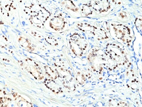
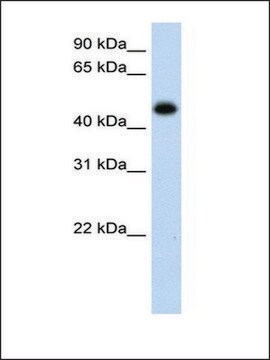
Anti-MUM1 antibody labels a 50kDa, multiple myeloma oncogene-1 (MUM1) protein. MUM1 is encoded by the MUM1/IRF-4 gene, which is mapped to 6q23-25 and identified as a myeloma-associated oncogene. It is a member of the interferon regulatory factor family of transcription factors and plays an important role in the regulation of gene expression in response to signaling by interferon and other cytokines. MUM1 positive cells express the protein in the nucleus in a diffuse and microgranular pattern. However, some positivity is also observed in the cytoplasm of MUM1-expressing cells. In normal/reactive lymphoid tissues, such as lymph node, this antibody stains plasma cells, some B-cells in the light zone of germinal centers, and a subset of T-cells (T-cells in germinal centers and interfollicular areas). MUM1 expression has been described in diffuse large B-cell lymphoma (DLBCL). Anti-MUM1 antibody can stain other B-cell lymphomas such as lymphoplasmacytic lymphoma, chronic lymphocytic leukemia, follicular lymphoma, marginal zone lymphoma, lymphoblastic lymphoma/leukemia, primary effusion lymphoma, DLBCL, Burkitt-like lymphoma, and classical Hodgkin lymphoma. However, the tumor cells in nodular lymphocyte predominant Hodgkin lymphoma are negative or only weakly positive. MUM1 is also expressed in plasma cell myeloma.

Qualité


IVD

IVD

IVD

RUO

Liaison

MUM1 Positive Control Slides, Product No. 358S, are available for immunohistochemistry (formalin-fixed, paraffin-embedded sections).

Forme physique

Solution in Tris Buffer, pH 7.3-7.7, with 1% BSA and <0.1% Sodium Azide.

Notes préparatoires

Download the IFU specific to your product lot and format
Note: This requires a keycode which can be found on your packaging or product label.

Download the latest released IFU
Note: This IFU may not apply to your specific product lot.

Autres remarques

For Technical Service please contact: 800-665-7284 or email: service@cellmarque.com

Informations légales

Cell Marque is a trademark of Merck KGaA, Darmstadt, Germany

Vous ne trouvez pas le bon produit ?  

Essayez notre Outil de sélection de produits.

Code de la classe de stockage

12 - Non Combustible Liquids

Classe de danger pour l'eau (WGK)

WGK 2

Point d'éclair (°F)

Not applicable

Point d'éclair (°C)

Not applicable


Certificats d'analyse (COA)

Recherchez un Certificats d'analyse (COA) en saisissant le numéro de lot du produit. Les numéros de lot figurent sur l'étiquette du produit après les mots "Lot" ou "Batch".

Déjà en possession de ce produit ?

Retrouvez la documentation relative aux produits que vous avez récemment achetés dans la Bibliothèque de documents.

Consulter la Bibliothèque de documents

W Kempf et al.
The British journal of dermatology, 158(6), 1280-1287 (2008-04-16)
Primary cutaneous CD30+ lymphoproliferative disorders include lymphomatoid papulosis (LyP) and primary cutaneous CD30+ anaplastic large T-cell lymphoma (ALCL). Because of overlapping histological features, it is impossible to distinguish ALCL from LyP on histological grounds. MUM1 (Multiple Myeloma oncogene 1) is
Antonino Carbone et al.
British journal of haematology, 117(2), 366-372 (2002-04-26)
Biological and clinical studies have shown that Hodgkin's disease (HD) can be divided into two major categories, termed nodular lymphocyte predominance HD (NLP HD) and classic HD (CHD). Within CHD four subtypes have been distinguished: nodular sclerosis, mixed cellularity, lymphocyte
Christine P Hans et al.
Blood, 103(1), 275-282 (2003-09-25)
Diffuse large B-cell lymphoma (DLBCL) can be divided into prognostically important subgroups with germinal center B-cell-like (GCB), activated B-cell-like (ABC), and type 3 gene expression profiles using a cDNA microarray. Tissue microarray (TMA) blocks were created from 152 cases of
Uma Sundram et al.
Modern pathology : an official journal of the United States and Canadian Academy of Pathology, Inc, 16(8), 802-810 (2003-08-16)
The diagnosis of malignant melanoma remains one of the most difficult to render in surgical pathology, partially because of its extreme histologic variability. Limits in the sensitivity and/or specificity of the currently available melanocytic markers such as anti-S100, HMB45, and

Notre équipe de scientifiques dispose d'une expérience dans tous les secteurs de la recherche, notamment en sciences de la vie, science des matériaux, synthèse chimique, chromatographie, analyse et dans de nombreux autres domaines..

Contacter notre Service technique