Accéder au contenu
Merck
Toutes les photos(1)

Key Documents

248M-1

Sigma-Aldrich

Ep-CAM/Epithelial Specific Antigen (MOC-31) Mouse Monoclonal Antibody

Se connecterpour consulter vos tarifs contractuels et ceux de votre entreprise/organisme


About This Item

Code UNSPSC :
12352203
Nomenclature NACRES :
NA.41

Source biologique

mouse

Niveau de qualité

100
500

Conjugué

unconjugated

Forme d'anticorps

culture supernatant

Type de produit anticorps

primary antibodies

Clone

MOC-31, monoclonal

Description

For In Vitro Diagnostic Use in Select Regions (See Chart)

Forme

buffered aqueous solution

Espèces réactives

human

Conditionnement

vial of 0.1 mL concentrate (248M-14)
vial of 0.5 mL concentrate (248M-15)
bottle of 1.0 mL predilute (248M-17)
vial of 1.0 mL concentrate (248M-16)
bottle of 7.0 mL predilute (248M-18)

Fabricant/nom de marque

Cell Marque

Technique(s)

immunohistochemistry (formalin-fixed, paraffin-embedded sections): 1:50-1:200

Isotype

IgG1κ

Contrôle

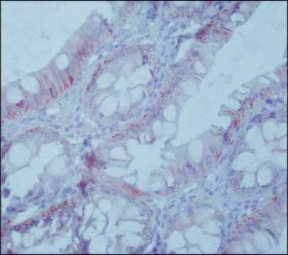
colon carcinoma

Conditions d'expédition

wet ice

Température de stockage

2-8°C

Visualisation

cytoplasmic

Informations sur le gène

human ... EPCAM(4072)

Description générale

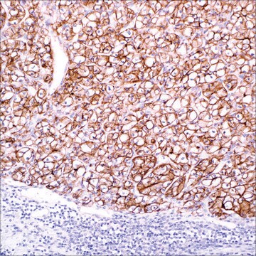
Epithelial cell adhesion molecule (Ep-CAM) is a transmembrane glycoprotein localized on the membrane of cells in most epithelial tissues. Immunoreactivity with the antibody to Ep-CAM has been seen in the majority of epithelial neoplasms, whereas most non-epithelial neoplasms do not show Ep-CAM expression. Ep-CAM is not expressed in mesothelial cells, hepatocytes, and lymphocytes. In conjunction with other markers, Ep-CAM can be used as an aid in determining neoplasms of epithelial origin, such as distinguishing between lung adenocarcinoma and mesothelioma.

Qualité


IVD

IVD

IVD

RUO

Liaison

Ep-CAM Positive Control Slides, Product No. 248S, are available for immunohistochemistry (formalin-fixed, paraffin-embedded sections).

Forme physique

Solution in Tris Buffer, pH 7.3-7.7, with 1% BSA and <0.1% Sodium Azide

Notes préparatoires

Download the IFU specific to your product lot and formatNote: This requires a keycode which can be found on your packaging or product label.

Autres remarques

For Technical Service please contact: 800-665-7284 or email: service@cellmarque.com

Informations légales

Cell Marque is a trademark of Merck KGaA, Darmstadt, Germany

Vous ne trouvez pas le bon produit ?  

Essayez notre Outil de sélection de produits.


Certificats d'analyse (COA)

Recherchez un Certificats d'analyse (COA) en saisissant le numéro de lot du produit. Les numéros de lot figurent sur l'étiquette du produit après les mots "Lot" ou "Batch".

Déjà en possession de ce produit ?

Retrouvez la documentation relative aux produits que vous avez récemment achetés dans la Bibliothèque de documents.

Consulter la Bibliothèque de documents

James S Lewis et al.
Modern pathology : an official journal of the United States and Canadian Academy of Pathology, Inc, 18(11), 1471-1481 (2005-06-25)
Sarcomatoid carcinomas are rare malignancies which represent poorly differentiated epithelial tumors that may be difficult to recognize as such. While some cases may have obvious epithelial areas, the sarcomatoid areas are poorly distinguishable from true sarcoma at the light microscopic
Murat Gokden et al.
Diagnostic cytopathology, 33(3), 166-172 (2005-08-04)
Differential diagnosis of primary and metastatic carcinomas of the liver can be problematic and may require immunohistochemical work-up. Recently, new immunohistochemical markers have been introduced with promising results in this area. We studied three of these markers, human hepatocyte antibody
Sanjay Kakar et al.
Archives of pathology & laboratory medicine, 131(11), 1648-1654 (2007-11-06)
Immunohistochemistry plays a crucial role in the diagnosis of hepatocellular carcinoma and in its distinction from other primary and metastatic neoplasms. Because limited tissue is available with fine-needle and core biopsies, appropriate selection of antibodies is imperative. To review the
Jonathan L Hecht et al.
Cancer, 108(1), 56-59 (2005-12-06)
It has been shown previously that detection of the epithelial membrane antigen using the mouse monoclonal antibody MOC-31 has diagnostic utility in the distinction between mesothelioma and metastatic carcinoma in body fluids. The current immunohistochemical study confirmed the effectiveness of
J E King et al.
Histopathology, 48(3), 223-232 (2006-01-25)
Immunohistochemistry is frequently employed to aid the distinction between mesothelioma and pulmonary adenocarcinoma metastatic to the pleura, but there is uncertainty as to which antibodies are most useful. We analysed published data in order to establish sensitivity and specificity of

Contenu apparenté

Diagnostic immunohistochemistry overview highlights its importance in cancer diagnosis and detecting infectious diseases in modern clinical pathology.

Notre équipe de scientifiques dispose d'une expérience dans tous les secteurs de la recherche, notamment en sciences de la vie, science des matériaux, synthèse chimique, chromatographie, analyse et dans de nombreux autres domaines..

Contacter notre Service technique