Description générale
Sterol regulatory element-binding protein 1 (UniProt: P36956; also known as SREBP-1, Class D basic helix-loop-helix protein 1, bHLHd1, Sterol regulatory element-binding transcription factor 1) is encoded by the SREBF1 (also known as BHLHD1, SREBP1) gene (Gene ID: 6720) in human. SREBP-1 is a multi-pass membrane protein found on nuclear, endoplasmic reticulum, and Golgi membranes. It attaches to membranes via two hydrophobic sequences (aa 477-497 and 536-556). It is expressed in a wide variety of tissues, but is most abundant in liver and adrenal gland. Six isoforms of SREBP-1 have been described that are produced by alternative splicing. Isoform SREBP-1C predominates in liver, adrenal gland, and ovary, whereas isoform SREBP-1A predominates in hepatoma cell lines. Both isoforms are found in kidney, brain, white fat, and muscle. It serves as a transcriptional activator that is essential for lipid homeostasis and regulates transcription of the LDL receptor gene. It is also involved in fatty acid and cholesterol synthesis. It acts by binds to the sterol regulatory element 1 (SRE-1) (5′-ATCACCCCAC-3′) and displays dual sequence specificity binding to both an E-box motif (5′-ATCACGTGA-3′) and to SRE-1 (5′-ATCACCCCAC-3′). SREBP-1 has two cytoplasmic domains (aa 1-487 and 569-1147), two transmembrane domains (aa 88-508 and 548-568), and one lumenal domain (aa 509-547). In the precursor form, which has a hairpin orientation in the membrane both the N-terminal transcription factor domain and the C-terminal regulatory domain face the cytoplasm. Two separate site-specific proteolytic cleavages are required for release of the transcriptionally active N-terminal domain. These cleavages are carried out by two distinct proteases, called site-1 protease (S1P; aa 490-491) and site-2 protease (S2P; aa 530-531). SREBP-1 can be phosphorylated by AMPK at Ser402, which suppress its processing and nuclear translocation, and thereby repress target gene expression.
Spécificité
Clone 2A4 is a mouse monoclonal antibody that detects Sterol regulatory element-binding protein 1. It targets an epitope within 107 amino acids from the N-terminal half.
Immunogène
His-tagged recombinant fragment corresponding to 107 amino acids from the N-terminal half of human Sterol regulatory element-binding protein 1 (SREBP-1).
Application
Anti-SREBP-1, clone 2A4, Cat. No. MABS2034, is a mouse monoclonal antibody that detects Sterol regulatory element-binding protein 1 and has been tested for use in Western Blotting.
Research Category
Signaling
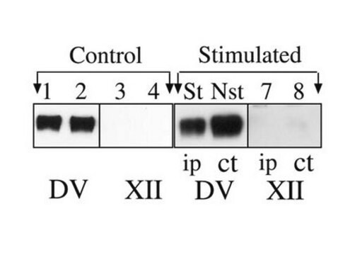
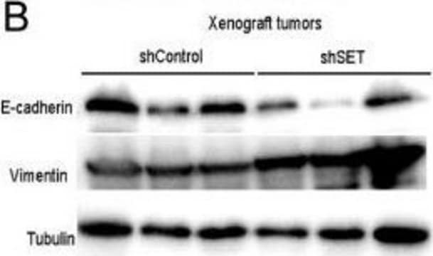
Western Blotting Analysis: A representative lot detected SREBP-1 in Western Blotting applications (Toth, J.I., et. al. (2004). Mol Cell Biol. 24(18):8288-300; Key, C.C., et. al. (2017). Cell Rep. 19(10):2116-2129; Rosenfeld, J.M., et. al. (1998). J Biol Chem. 273(26):16112-21; Sato, R., et. al. (1994). J Biol Chem. 269(25):17267-73).
Qualité
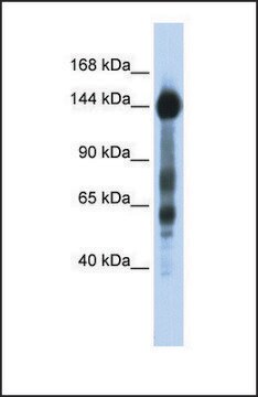
Evaluated by Western Blotting in HeLa cell lysate.
Western Blotting Analysis: 1 µg/mL of this antibody detected SREBP-1 in HeLa cell lysate.
Description de la cible
~135 kDa observed; 121.68 kDa calculated. Uncharacterized bands may be observed in some lysate(s).
Forme physique
Format: Purified
Protein G purified
Purified mouse monoclonal antibody IgG1 in buffer containing 0.1 M Tris-Glycine (pH 7.4), 150 mM NaCl with 0.05% sodium azide.
Stockage et stabilité
Stable for 1 year at 2-8°C from date of receipt.
Autres remarques
Concentration: Please refer to lot specific datasheet.
Clause de non-responsabilité
Unless otherwise stated in our catalog or other company documentation accompanying the product(s), our products are intended for research use only and are not to be used for any other purpose, which includes but is not limited to, unauthorized commercial uses, in vitro diagnostic uses, ex vivo or in vivo therapeutic uses or any type of consumption or application to humans or animals.
