Accéder au contenu
Merck
Toutes les photos(2)

Principaux documents

ABS535

Sigma-Aldrich

Anti-Arginase-1 Antibody

from chicken

Synonyme(s) :

Type 1 Arginase

ARG1

Se connecterpour consulter vos tarifs contractuels et ceux de votre entreprise/organisme


About This Item

Code UNSPSC :
12352203
eCl@ss :
32160702
Nomenclature NACRES :
NA.41

Source biologique

chicken

Niveau de qualité

Forme d'anticorps

purified immunoglobulin

Type de produit anticorps

primary antibodies

Clone

polyclonal

Espèces réactives

guinea pig, human, mouse, rat

Technique(s)

immunohistochemistry: suitable
western blot: suitable

Isotype

IgY

Numéro d'accès NCBI

Numéro d'accès UniProt

Conditions d'expédition

wet ice

Modification post-traductionnelle de la cible

unmodified

Informations sur le gène

human ... ARG1(383)
mouse ... Arg1(11846)
rat ... Arg1(29221)

Description générale

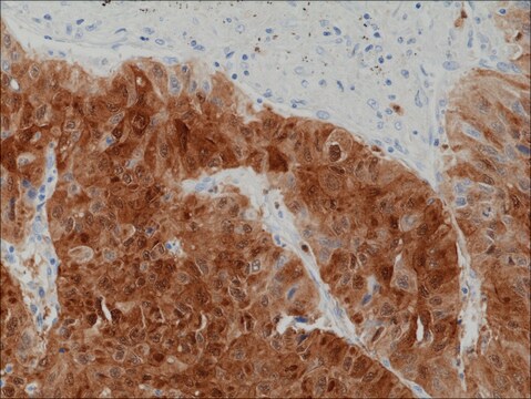
Arginase-1, the protein encoded by the ARG1 gene, is the enzyme that catalyzes the breakdown of L-arginine into ornithine and urea. ARG1 is a key player in the Urea cycle. There are two distinct isoforms of Arginase, Arginase I and Arginase II. Arginase I (also called the liver type arginase or Type 1 arginase) is a highly expressed cytosolic enzyme in the liver and other tissues, including the brain. Arginase II or kidney-type arginase is also widely expressed at low levels in many tissues but localized to the mitochondria primarily. Interestingly, Arginase 1 has neuroprotective properties. Arginase I is essential for protecting motor neurons from nitric oxide dependent apoptosis that can be induced upon cellular stress or trophic factor removal. By suppressing nitric oxide production, Arginase prevents the formation of peroxynitrite and its subsequent cellular toxicity. Furthermore, Arginase 1 appears to play critical roles throughout the CNS for survival and regeneration of neurons and axons. In cancer, Arginase 1, already a useful marker for hepatocytes is also an excellent marker for hepatocellular cancers.

Immunogène

Full-length recombinant protein corresponding to Rat Arginase-1.

Application

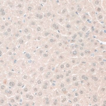
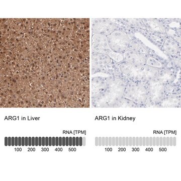
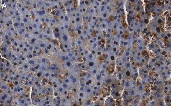
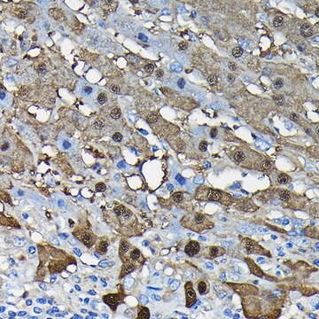
Analyse par immunohistochimie : A representative lot detected Arginase-1 in bronchoalveolar lavage cells (Zimmermann, N., et al. (2003). –
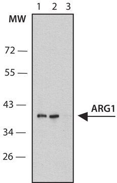
A representative lot detected Arginase-1 in RAW cell lysate (Morris, S., et al. (1998). Am J Physiol Endocrinol Metab 275:E740-E747, 1998).
Domaine de recherche
Signalisation
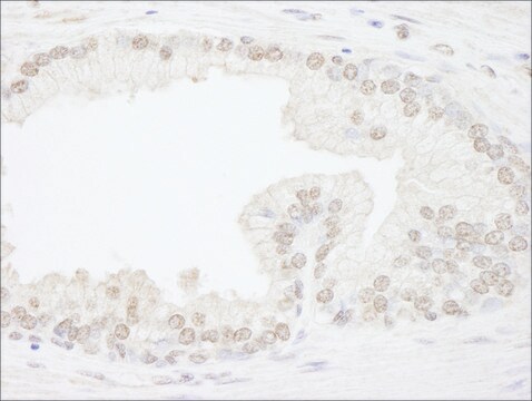
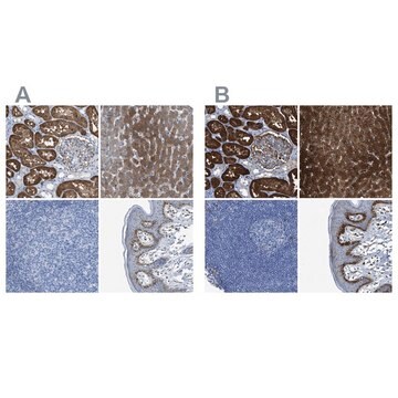
This Anti-Arginase-1 Antibody is validated for use in Western Blotting and Immunohistochemistry for the detection of Arginase-1.

Qualité


µµ

Description de la cible

Poids réel (observé) : env. 38 kDa

Forme physique

Format : Produit purifié
IgY purified from Yolk
Purified chicken Polyclonal other in buffer containing PBS with 0.05% sodium azide.

Stockage et stabilité

Stable entre 2 et 8 °C pendant 1 an à compter de la date de réception.

Autres remarques

Concentration : Voir la fiche technique du lot concerné.

Clause de non-responsabilité

Sauf indication contraire dans notre catalogue ou toute autre documentation associée au(x) produit(s), nos produits sont uniquement destinés à la recherche et ne sauraient être utilisés à d'autres fins, ce qui inclut, sans s'y limiter, les utilisations commerciales non autorisées, les utilisations diagnostiques in vitro, les utilisations thérapeutiques ex vivo ou in vivo, ou tout type de consommation ou d'application chez l'être humain ou chez l'animal.

Vous ne trouvez pas le bon produit ?  

Essayez notre Outil de sélection de produits.

Code de la classe de stockage

12 - Non Combustible Liquids

Classe de danger pour l'eau (WGK)

WGK 2

Point d'éclair (°F)

Not applicable

Point d'éclair (°C)

Not applicable


Certificats d'analyse (COA)

Recherchez un Certificats d'analyse (COA) en saisissant le numéro de lot du produit. Les numéros de lot figurent sur l'étiquette du produit après les mots "Lot" ou "Batch".

Déjà en possession de ce produit ?

Retrouvez la documentation relative aux produits que vous avez récemment achetés dans la Bibliothèque de documents.

Consulter la Bibliothèque de documents

Tim Tian Y Han et al.
Frontiers in bioengineering and biotechnology, 9, 642465-642465 (2021-04-06)
Cell-based therapies involving the delivery of adipose-derived stromal cells (ASCs) on decellularized adipose tissue (DAT) scaffolds are a promising approach for soft tissue augmentation and reconstruction. Our lab has recently shown that culturing human ASCs on DAT scaffolds within a
Nicolas De Vos et al.
Clinics and research in hepatology and gastroenterology, 45(3), 101478-101478 (2020-07-06)
Preoperative imaging and histopathology, immunohistochemistry and molecular analysis after resection of 2 hepatocellular adenomas (HCAs) (20 and 2cm) in a 53-year-old female patient were performed. On imaging, the large lesion resembled a myxoid HCA, while the small lesion resembled a
Asiyeh Golabchi et al.
Biomaterials, 180, 225-239 (2018-07-28)
The chronic performance of implantable neural electrodes is hindered by inflammatory brain tissue responses, including microglia activation, glial scarring, and neuronal loss. Melatonin (MT) has shown remarkable neuroprotective and neurorestorative effects in treating central nervous system (CNS) injuries and degeneration
Julia S Brunner et al.
Nature communications, 11(1), 431-431 (2020-01-24)
Multinucleated giant cells (MGCs) are implicated in many diseases including schistosomiasis, sarcoidosis and arthritis. MGC generation is energy intensive to enforce membrane fusion and cytoplasmic expansion. Using receptor activator of nuclear factor kappa-Β ligand (RANKL) induced osteoclastogenesis to model MGC
Matthew J Kan et al.
The Journal of neuroscience : the official journal of the Society for Neuroscience, 35(15), 5969-5982 (2015-04-17)
The pathogenesis of Alzheimer's disease (AD) is a critical unsolved question; and although recent studies have demonstrated a strong association between altered brain immune responses and disease progression, the mechanistic cause of neuronal dysfunction and death is unknown. We have

Notre équipe de scientifiques dispose d'une expérience dans tous les secteurs de la recherche, notamment en sciences de la vie, science des matériaux, synthèse chimique, chromatographie, analyse et dans de nombreux autres domaines..

Contacter notre Service technique