Accéder au contenu
Merck
Toutes les photos(1)

Principaux documents

05-629-IM

Sigma-Aldrich

Anti-Histone H1o/H5 Antibody, clone 3H9

ascites fluid, clone 3H9, from mouse

Synonyme(s) :

Histone H5, Histone H1o

Se connecterpour consulter vos tarifs contractuels et ceux de votre entreprise/organisme


About This Item

Code UNSPSC :
12352203
eCl@ss :
32160702
Nomenclature NACRES :
NA.41

Source biologique

mouse

Niveau de qualité

Forme d'anticorps

ascites fluid

Type de produit anticorps

primary antibodies

Clone

3H9, monoclonal

Espèces réactives

human, chicken

Conditionnement

antibody small pack of 25 μL

Technique(s)

ChIP: suitable (ChIP-chip)
flow cytometry: suitable
immunocytochemistry: suitable
immunofluorescence: suitable
western blot: suitable

Isotype

IgG2aκ

Numéro d'accès NCBI

Numéro d'accès UniProt

Modification post-traductionnelle de la cible

unmodified

Informations sur le gène

human ... H1-0(3005)

Description générale

Histone H1/H5 (UniProt: P02259) are linker histones that are involved in the organization and maintenance of chromatin higher order structure and they play an active role in the control of DNA replication and cell proliferation. Linker histones are essential for chromatin filament formation, and they play key roles in the regulation of gene expression. Histone H1o is a histone H5-like protein found in mammalian species. It is present in the linker region of chromatin and replaces H1 there. Up to one third of the H1 can be replaced in some tissues. Histone H5 performs the same function as H1, being necessary for the condensation of nucleosome chains into higher order structures, and replaces histone H1 in certain cells. Histone H5 is found in erythroid cells in avian species. Histone H5 consists of a central globular domain (GH5) that is essential for nucleosome binding and is flanked by basic N- and C-terminal tails. It is reported that binding of either histone H5 or GH5 to nucleosomes protects an additional 20 bp of linker DNA from micrococcal nuclease digestion. Globular domain of linker histone H5 (GH5) contains three distinct DNA-binding sites, through which GH5 contacts the DNA at the nucleosome dyad and the linker DNA strands entering and exiting the nucleosome. (Ref.: Segers, A et al (1991). J. Biol. Chem. 266(3); 1502-1508; Fan, L., and Roberts, VA (2006). Proc. Natl. Acad. Sci. USA. 113 (22); 8384 8389; Smith, BH and Jones, EW (1980). Nucleic Acids Res. 8(24); 6069-6079).

Spécificité

Clone 3H9 detects Histone H1o/H5 in human and chicken species. it targets an epitope within 46 amino acids from the N-terminal region.

Immunogène

Native histone H5 protein purified from chicken erythrocytes.

Application

Anti-Histone H1o/H5, clone 3H9 , Cat. No. 05-629-I, is a highly specific mouse monoclonal antibody that targets Histone H5 and has been tested in Chromatin Immunoprecipitation (ChIP), Flow Cytometry, Immunocytochemistry, Immunofluorescence, and Western Blotting.
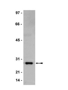
Western Blotting Analysis: A representative lot detected Histone H1o/H5 in Western Blotting applications (Torres, C.M., et. al. (2016). Science. 353(6307)).

Chromatin Immunoprecipitation Analysis: A representative lot detected Histone H1o/H5 in Chromatin Immunoprecipitation applications (Torres, C.M., et. al. (2016). Science. 353(6307)).

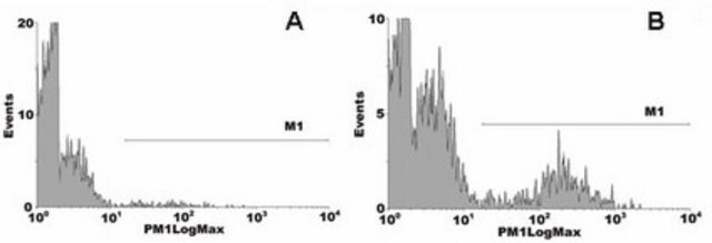
Flow Cytometry Analysis: A representative lot detected Histone H1o/H5 in Flow Cytometry applications (Torres, C.M., et. al. (2016). Science. 353(6307)).

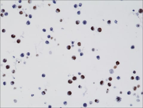
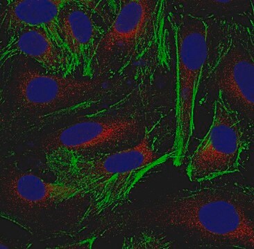
Immunocytochemistry Analysis: A representative lot detected Histone H1o/H5 in Immunocytochemistry applications (Torres, C.M., et. al. (2016). Science. 353(6307)).

Immunofluorescence Analysis: A representative lot detected Histone H1o/H5 in Immunofluorescence applications (Torres, C.M., et. al. (2016). Science. 353(6307)).

Qualité

Evaluated by Western Blotting in HeLa cells acid extract.

Western Blotting Analysis: A 1:20,000 dilution of this antibody detected Histone H1o in 10 µg of HeLa cells acid extract.

Description de la cible

~30 kDa observed; 20.73 kDa calculated. Uncharacterized bands may be observed in some lysate(s).

Forme physique

Mouse monoclonal antibody in ascites without preservatives.

Autres remarques

Concentration: Please refer to lot specific datasheet.

Vous ne trouvez pas le bon produit ?  

Essayez notre Outil de sélection de produits.

Code de la classe de stockage

10 - Combustible liquids

Classe de danger pour l'eau (WGK)

WGK 1

Point d'éclair (°F)

Not applicable

Point d'éclair (°C)

Not applicable


Certificats d'analyse (COA)

Recherchez un Certificats d'analyse (COA) en saisissant le numéro de lot du produit. Les numéros de lot figurent sur l'étiquette du produit après les mots "Lot" ou "Batch".

Déjà en possession de ce produit ?

Retrouvez la documentation relative aux produits que vous avez récemment achetés dans la Bibliothèque de documents.

Consulter la Bibliothèque de documents

Notre équipe de scientifiques dispose d'une expérience dans tous les secteurs de la recherche, notamment en sciences de la vie, science des matériaux, synthèse chimique, chromatographie, analyse et dans de nombreux autres domaines..

Contacter notre Service technique