
R5403
Monoclonal Anti-Rhodopsin antibody produced in mouse
clone 1D4, purified immunoglobulin, buffered aqueous solution
Synonym(s):
Anti-CSNBAD1, Anti-OPN2, Anti-RP4
About This Item
Recommended Products

biological source
mouse
Quality Level
conjugate
unconjugated
antibody form
purified immunoglobulin
antibody product type
primary antibodies
clone
1D4, monoclonal
form
buffered aqueous solution
species reactivity
human, rat, bovine
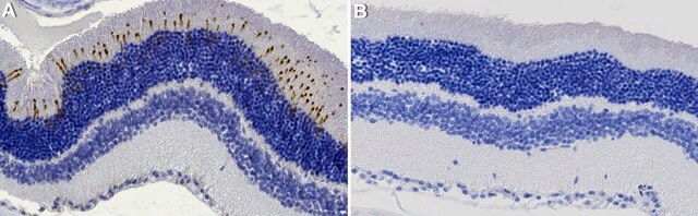
technique(s)
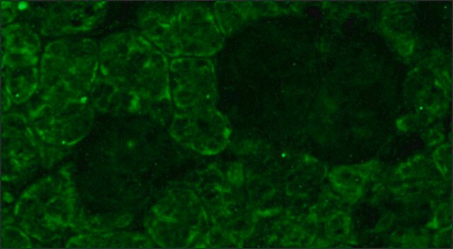
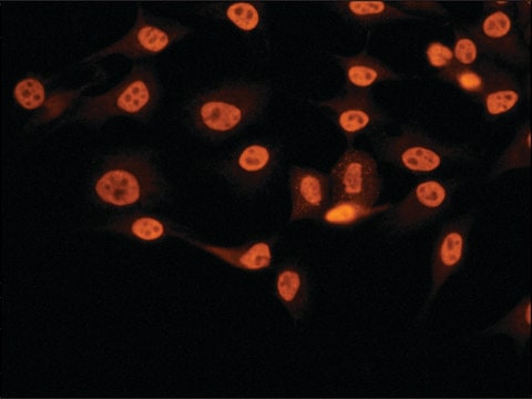
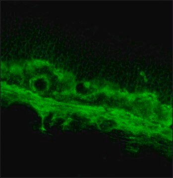
immunocytochemistry: 1:1,000 using human retinal samples
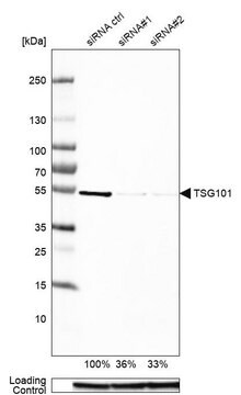
western blot: 1:1,000 using Sf9 cells expressing the bovine gene
isotype
IgG1
UniProt accession no.
shipped in
wet ice
storage temp.
−20°C
target post-translational modification
unmodified
Gene Information
human ... RHO(6010)
rat ... Rho(24717)
General description
Specificity
Immunogen
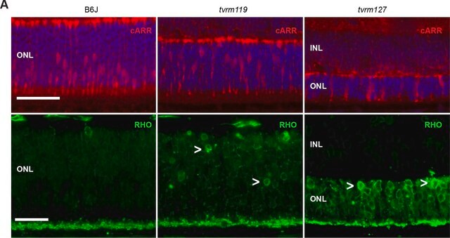
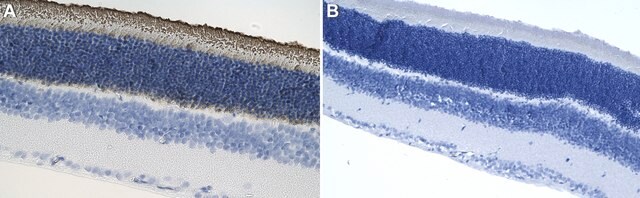
Application
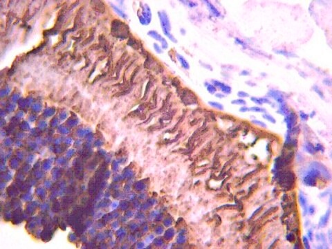
Immunohistochemistry (1 paper)
Physical form
Disclaimer
Not finding the right product?
Try our Product Selector Tool.
recommended
Storage Class Code
10 - Combustible liquids
WGK
nwg
Flash Point(F)
Not applicable
Flash Point(C)
Not applicable
Choose from one of the most recent versions:
Already Own This Product?
Find documentation for the products that you have recently purchased in the Document Library.
Our team of scientists has experience in all areas of research including Life Science, Material Science, Chemical Synthesis, Chromatography, Analytical and many others.
Contact Technical Service