Skip to Content
Merck
All Photos(1)

Key Documents

G5421

Sigma-Aldrich

Monoclonal Anti-Granulocyte Colony Stimulating Factor antibody produced in rat

clone 67604, purified immunoglobulin, lyophilized powder

Synonym(s):

Anti-Csf3, Anti-Csfg, Anti-G-CSF, Anti-MGI-IG

Sign Into View Organizational & Contract Pricing


About This Item

UNSPSC Code:
51111800
NACRES:
NA.41

biological source

rat

Quality Level

conjugate

unconjugated

antibody form

purified immunoglobulin

antibody product type

primary antibodies

clone

67604, monoclonal

form

lyophilized powder

species reactivity

mouse

technique(s)

capture ELISA: 2-8 μg/mL
neutralization: suitable
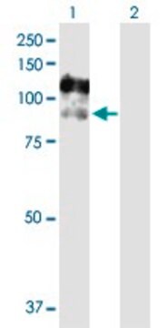
western blot: 1-2 μg/mL

isotype

IgG1

UniProt accession no.

storage temp.

−20°C

Gene Information

mouse ... Csf3(12985)

General description

Mature myeloid cells are derived from bone-marrow derived precursor cells by the activity of three colony stimulating factors, granulocyte/macrophage colony-stimulating factor (GM-CSF), macrophage colony-stimulating factor (M-CSF) and granulocyte colony-stimulating factor (G-CSF). The expression of G-CSF is the result of activation of pathways in response to tumor necrosis factor-α, LPS and toll-like receptor ligands. The pathways that induce the effects of G-CSF are PI3K, ERK1/2 and p38. The generation of neutrophils and induction of the circulation of hematopoietic stem cells from the bone marrow are the main functions of G-CSF. There are additional activities attributed to G-CSF such as neuroprotection, immunomodulation and generation of cardiac cells and dendritic cells. Large quantities of G-CSF are secreted in a variety of myeloid and non-myeloid cancers and rheumatoid arthritis
Monoclonal anti-granulocyte colony stimulating factor recognizes mouse G-CSF. The antibody shows less than 0.06% cross-reactivity with recombinant human G-CSF.

Immunogen

purified, recombinant mouse granulocyte colony stimulating factor, expressed in E. coli.

Application

Anti-granulocyte colony stimulating factor antibody may be used for immunoblotting at a working concentration of 1-2 μg/ml. For ELISA the recommended concentration range is 2-8 μg/ml. The antibody is also suitable for neutralization assays.

Physical form

Lyophilized from a 0.2 μm filtered solution in phosphate buffered saline with 5% trehalose.

Disclaimer

Unless otherwise stated in our catalog or other company documentation accompanying the product(s), our products are intended for research use only and are not to be used for any other purpose, which includes but is not limited to, unauthorized commercial uses, in vitro diagnostic uses, ex vivo or in vivo therapeutic uses or any type of consumption or application to humans or animals.

Not finding the right product?  

Try our Product Selector Tool.

Storage Class Code

13 - Non Combustible Solids

WGK

WGK 1

Flash Point(F)

Not applicable

Flash Point(C)

Not applicable

Personal Protective Equipment

dust mask type N95 (US), Eyeshields, Gloves

Choose from one of the most recent versions:

Certificates of Analysis (COA)

Lot/Batch Number

Don't see the Right Version?

If you require a particular version, you can look up a specific certificate by the Lot or Batch number.

Already Own This Product?

Find documentation for the products that you have recently purchased in the Document Library.

Visit the Document Library

Lorraine Pelosof et al.
Epigenetics, 12(7), 540-550 (2016-12-06)
Epigenetic control of gene expression is a major determinant of tumor phenotype and has been found to influence sensitivity to individual chemotherapeutic agents. Glutathione peroxidase 3 (GPX3, plasma glutathione peroxidase) is a key component of cellular antioxidant regulation and its
F Liu et al.
Immunity, 5(5), 491-501 (1996-11-01)
We have generated mice carrying a homozygous null mutation in the granulocyte colony-stimulating factor receptor (G-CSFR) gene. G-CSFR-deficient mice have decreased numbers of phenotypically normal circulating neutrophils. Hematopoietic progenitors are decreased in the bone marrow, and the expansion and terminal
R R Ji et al.
Nature neuroscience, 2(12), 1114-1119 (1999-11-26)
We investigated the involvement of extracellular signal-regulated protein kinases (ERK) within spinal neurons in producing pain hypersensitivity. Within a minute of an intense noxious peripheral or C-fiber electrical stimulus, many phosphoERK-positive neurons were observed, most predominantly in lamina I and
Sebastian Stösser et al.
Journal of molecular medicine (Berlin, Germany), 89(4), 321-329 (2010-11-17)
A variety of cancers are accompanied by debilitating pain, which constitutes the primary reason for poor quality of life in cancer patients. There is an urgent demand for the development of specific mechanism-based therapies against cancer pain. Recently, important advances
Sophie Pezet et al.
The European journal of neuroscience, 21(7), 1785-1797 (2005-05-05)
The serine/threonine kinase Akt/PKB has been implicated in cell survival signalling in many cell types, including the dorsal root ganglion (DRG). However, little is known about its role in physiological and pathophysiological conditions in the adult sensory and nociceptive system.

Our team of scientists has experience in all areas of research including Life Science, Material Science, Chemical Synthesis, Chromatography, Analytical and many others.

Contact Technical Service