B3111
ANTI-FLAG® M2 antibody, Mouse monoclonal
Clone M2, purified from hybridoma cell culture in bioreactor
Synonym(s):
Anti-ddddk, Anti-dykddddk, M2 clone ANTI-FLAG
Sign Into View Organizational & Contract Pricing
All Photos(7)
About This Item
UNSPSC Code:
12352203
NACRES:
NA.43
Recommended Products
biological source
mouse
antibody form
purified immunoglobulin (purified IgG1 subclass)
clone
M2, monoclonal
shelf life
4 yr
purified by
using Protein A
storage temp.
−20°C
General description
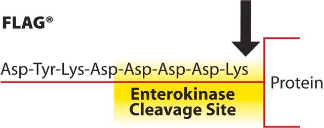
Monoclonal ANTI-FLAG M2 is a purified immunoglobulin, IgG1, monoclonal antibody, purified from culture supernatant of hybridoma cells, that binds to FLAG® fusion proteins. Unlike ANTI-FLAG M1 antibody, the M2 antibody will recognize the FLAG sequence at the N-terminus, Met-N-terminus, C-terminus, or at an internal site of FLAG fusion proteins. Monoclonal ANTI-FLAG M2 is useful for identification and capture of FLAG fusion proteins by common immunological procedures such as Western blots and immunoprecipitation. It is also useful for affinity purification of FLAG fusion proteins when bound to a solid support.
form: solution pH 7.4, containing 15 mM sodium azide
concentration: 3.0-5.0 mg/mL
form: solution pH 7.4, containing 15 mM sodium azide
concentration: 3.0-5.0 mg/mL
Application
IB, IF, IP, FACS, ELISA
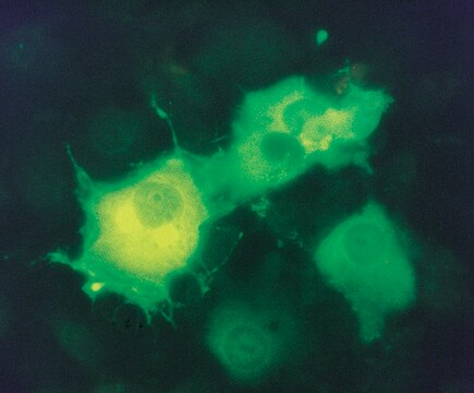
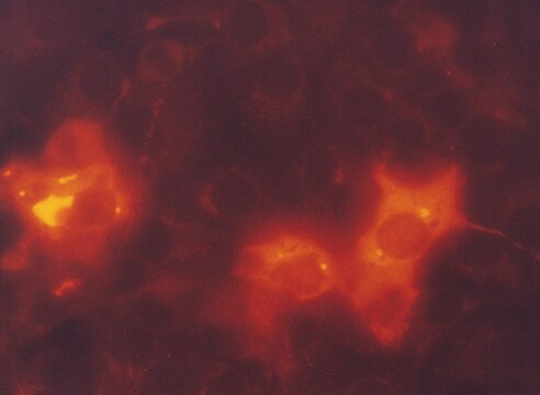
Antibody is recommended for use in several applications such as immunoblotting, immunoprecipitation, immunofluorescence, flow cytometry, and ELISA.
Learn more product details in our FLAG® application portal.
Antibody is recommended for use in several applications such as immunoblotting, immunoprecipitation, immunofluorescence, flow cytometry, and ELISA.
Learn more product details in our FLAG® application portal.
Packaging
polypropylene screw cap vial
Preparation Note
Dilute the antibody solution from 0.5-10 ug/mL in specified buffer
Legal Information
ANTI-FLAG is a registered trademark of Merck KGaA, Darmstadt, Germany
FLAG is a registered trademark of Merck KGaA, Darmstadt, Germany
Storage Class Code
12 - Non Combustible Liquids
WGK
nwg
Flash Point(F)
Not applicable
Flash Point(C)
Not applicable
Choose from one of the most recent versions:
Certificates of Analysis (COA)
Lot/Batch Number
Sorry, we don't have COAs for this product available online at this time.
If you need assistance, please contact Customer Support.
Already Own This Product?
Find documentation for the products that you have recently purchased in the Document Library.
Yi-Min Chu et al.
Frontiers in oncology, 12, 900166-900166 (2022-10-04)
DLC1 (deleted in liver cancer-1) is downregulated or deleted in colorectal cancer (CRC) tissues and functions as a potent tumor suppressor, but the underlying molecular mechanism remains elusive. We found that the conditioned medium (CM) collected from DLC1-overexpressed SW1116 cells
Yanchen Ma et al.
Glia, 70(2), 379-392 (2021-11-02)
Myelin sheath is an important structure to maintain functions of the nerves in central nervous system. Protein palmitoylation has been established as a sorting determinant for the transport of myelin-forming proteins to the myelin membrane, however, its function in the
Jiajia Zhang et al.
Cancer letters, 501, 43-54 (2020-12-29)
TP53 binding protein 1 (53BP1) plays an important role in DNA damage repair and maintaining genomic stability. However, the mutations of 53BP1 in human cancers have not been systematically examined. Here, we have analyzed 541 somatic mutations of 53BP1 across
Maria Jesús García-Murria et al.
Nature communications, 11(1), 6056-6056 (2020-11-29)
Viral control of programmed cell death relies in part on the expression of viral analogs of the B-cell lymphoma 2 (Bcl2) protein known as viral Bcl2s (vBcl2s). vBcl2s control apoptosis by interacting with host pro- and anti-apoptotic members of the
Silvia Martini et al.
Nature communications, 12(1), 6934-6934 (2021-11-28)
The PKCε-regulated genome protective pathway provides transformed cells a failsafe to successfully complete mitosis. Despite the necessary role for Aurora B in this programme, it is unclear whether its requirement is sufficient or if other PKCε cell cycle targets are
Our team of scientists has experience in all areas of research including Life Science, Material Science, Chemical Synthesis, Chromatography, Analytical and many others.
Contact Technical Service


