A1729
Activin B human
≥90% (SDS-PAGE), recombinant, expressed in CHO cells, lyophilized, suitable for cell culture
Sign Into View Organizational & Contract Pricing
All Photos(1)
About This Item
Recommended Products
Product Name
Activin B human, recombinant, expressed in CHO cells, suitable for cell culture
biological source
human
Quality Level
recombinant
expressed in CHO cells
Assay
≥90% (SDS-PAGE)
form
lyophilized
potency
0.30-1.50 ng/mL ED50
mol wt
calculated mol wt ~14.5 kDa
packaging
pkg of 5 μg
storage condition
avoid repeated freeze/thaw cycles
technique(s)
cell culture | mammalian: suitable
impurities
endotoxin, tested
storage temp.
−20°C
Biochem/physiol Actions
Activin-B is involved in the regulation of a variety of cell functions that overlap the actions of activin-A; however, activin-B also regulates many processes that are distinct from activin-A activities. The functional differences between activin-A and activin-B have recently been reviewed, Thompson TB, et al. (2004).
Similar to activin-A, activin-B modulates follicle stimulating hormone (FSH) secretion and hemoglobin synthesis, DePaolo LV, et al. (1992); Mason AJ, et al. (1989). It is involved in regulation of the menstrual cycle, Liu J, et al. (2001).
Activin-B is not essential for embryo development or survival, but it does have important roles in development. It stimulates spermatogonial proliferation, Mather JP, et al. (1990) and is localized in specific cells, gonocytes and interstitial Leydig cells, and tissues, rete testis and epididymal epithelium, associated with human testis duct system development, Anderson RA, et al. (2002). Activin-B is found in follicle cells surrounding oocytes, Dohrmann CE, et al. (1993) and been shown to increase the rate of oocytes maturation in a dose and time dependent manner, Pang Y and Ge W. (1999, 2002). It is required for successful mammogenesis leading to lactation, Robinson GW and Hennighausen L. (1997). Recent studies suggest that activin-B may regulate adipocyte differentiation, Kogame, M, et al. (2006).
Activin-B has been linked to several aspects of embryo development. Activin-B is first detected in blastula-stage embryos, Thomsen G, et al. (1990) after midblastula transition and homogeneously distributed during blastula and early gastrula stages. It becomes restricted to the dorso-anterior region in neurula-stage embryos and by early tailbud stage it is restricted to brain, eye anlagen, visceral pouches, otic vesicles and the anterior notochord, Dohrmann CE, et al. (1993). Activin-A and -B localizations are different in 3.5 and 4.5 day mouse blastocyst, Paulusman CC, et al. (1994).
Nakamura, T et al. (1992) suggested that the primary function of Activin-B may involve mesoderm-inducing activity and early development modulation. Schrewe H, et al (1994) and Vassalli A, et al. (1994), reported that activin-B was not essential for survival or for mesoderm formation (in mouse), but that it played a role in late fetal development and female fecundity. A role for activin-B in axial formation was suspected because ectopic expression produced second body axis embryos, Thomsen G, et al. (1990), Mitrani E, et al. (1990).
The roles of activin-B during embryo development are starting to emerge. Activin-B may modulate gap junction permeability in embryos, Olson DJ and Moon RT. (1992). Activin-B has been shown to modulate the morphogenesis of the roof plate (RP) of midbrain wherein it inhibits roof plate differentiation, Alexandre P, et al. (2006). Activin-B signals cell cycle arrest in cells of the involuting dorsal axial mesoderm, Ramis JM, et al. (2007).
Activin-B is involved in the development of the adrenal gland and pancreas. It is present in normal adrenal medulla, but absent in the cortex, Salmenkivi K et al. (2001). Interestingly, Activin-B may be a marker for benign adrenal pheochromocytomas, Salmenkivi K et al. (2001).
The role of activin-B in pancreas development is of particular interest because of efforts to use human embryonic stem cells (hESC) as precursors to make insulin producing cells for treatment of diabetes. Activin-B has been shown to promote expression of the pancreas marker Pdx1 gene in cells of differentiated embryoid bodies (EB), in culture, Frandsen U, et al. (2007). Most recently, Jafary H, et al. (2008) induced insulin-secreting cells for ES by adding activin-B to nestin-positive selection protocol cell.
Activins have a wide range of biological activities including mesoderm induction, neural cell differentiation, bone remodeling, hematopoiesis, and reproductive physiology. Activins influence erythropoiesis and the potentiation of erythroid colony formation, oxytocin secretion, paracrine, and autocrine regulation.
Physical form
Lyophilized from a 0.2 μm filtered solution in 30% acetonitrile and 0.1% TFA containing 0.25 mg bovine serum albumin.
Analysis Note
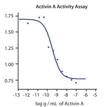
The biological activity is measured by its ability to induce hemoglobin expression in K562 cells.
Signal Word
Warning
Hazard Statements
Precautionary Statements
Hazard Classifications
Acute Tox. 4 Inhalation - Eye Irrit. 2
Storage Class Code
3 - Flammable liquids
WGK
WGK 2
Flash Point(F)
Not applicable
Flash Point(C)
Not applicable
Personal Protective Equipment
dust mask type N95 (US), Eyeshields, Gloves
Choose from one of the most recent versions:
Already Own This Product?
Find documentation for the products that you have recently purchased in the Document Library.
Erythroid differentiation bioassays for activin.
R H Schwall et al.
Methods in enzymology, 198, 340-346 (1991-01-01)
A J Mason et al.
Biochemical and biophysical research communications, 135(3), 957-964 (1986-03-28)
The complete amino acid sequences of two forms of human ovarian inhibin have been determined through cloning and nucleotide sequencing of cDNAs encoding their individual subunit precursors. The alpha subunit common to both forms of human inhibin is homologous (84
Our team of scientists has experience in all areas of research including Life Science, Material Science, Chemical Synthesis, Chromatography, Analytical and many others.
Contact Technical Service


