Direkt zum Inhalt
Merck

C5992

Sigma-Aldrich

Monoklonales Anti-pan-Zytokeratin

clone PCK-26, purified from hybridoma cell culture

Synonym(e):

Monoclonal Anti-pan-Cytokeratin, Anti-Cytokeratin, Anti-Cytokeratin - Monoclonal Anti-Cytokeratin, pan antibody produced in mouse

Anmeldenzur Ansicht organisationsspezifischer und vertraglich vereinbarter Preise


About This Item

UNSPSC-Code:
12352203
NACRES:
NA.41

Biologische Quelle

mouse

Qualitätsniveau

Konjugat

unconjugated

Antikörperform

purified from hybridoma cell culture

Antikörper-Produkttyp

primary antibodies

Klon

PCK-26, monoclonal

Form

buffered aqueous solution

Speziesreaktivität

human, chicken, snake, hamster, pig, goat, feline, bovine, carp, rat, rabbit, canine, lizard, sheep, mouse, guinea pig

Verpackung

antibody small pack of 25 μL

Konzentration

~1.5 mg/mL

Methode(n)

dot blot: suitable
immunocytochemistry: suitable
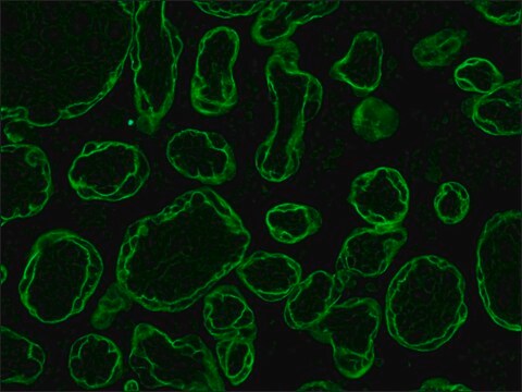
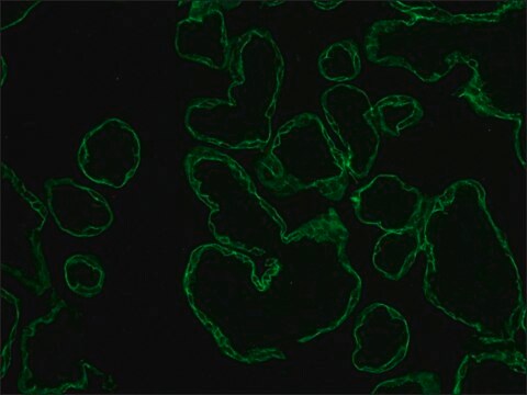
immunohistochemistry: 10-20 μg/mL using human placenta or skin
western blot: suitable

Isotyp

IgG1

Versandbedingung

dry ice

Lagertemp.

−20°C

Posttranslationale Modifikation Target

unmodified

Suchen Sie nach ähnlichen Produkten? Aufrufen Leitfaden zum Produktvergleich

Allgemeine Beschreibung

Monoclonal Anti-Pan Cytokeratin (mouse IgG1 isotype) is derived from the PCK-26 hybridoma produced by the fusion of mouse myeloma cells and splenocytes from BALB/c mice immunized with a cytokeratin preparation from human epidermis. Cytokeratins are a group of at least 29 different proteins. They are characteristic of epithelial and trichocytic cells. Cytokeratin peptide 1 (68 kDa) is expressed together with cytokeratin 10 in the suprabasal cell layers or the differentiation compartment of the epidermis. Its expression increases with epidermal maturation and is modified post translationally in terminally differentiated keratinocytes of the stratum corneum. Cytokeratin peptide 5 (58 kDa) is the primary type II keratin in stratified epithelia, while cytokeratin type 8 (52 kDa) is a major type II keratin in simple epithelia. Cytokeratin 6 (56 kDa) is a "hyperproliferation" cytokeratin expressed in tissues with natural or pathological high turnover. Monoclonal antibodies to cytokeratins are specific markers of epithelial cell differentiation and have been widely used as tools in tumor identification and classification.

Spezifität

Der Antikörper erkennt ein Epitop auf Typ-II-Cytokeratinen 1, 5, 6 und 8. PCK-26 ist ein Breitspektrum-Antikörper, der spezifisch mit einer Vielfalt an normalen, reaktiven und neoplastischen Epithelgeweben reagiert. Der Antikörper reagiert mit einfachen, verhornten und unverhornten Plattenepitheln und pseudostratifizierten Epitheln.

Immunogen

Cytokeratin aus der humanen Epidermis.

Anwendung

Applications in which this antibody has been used successfully, and the associated peer-reviewed papers, are given below.
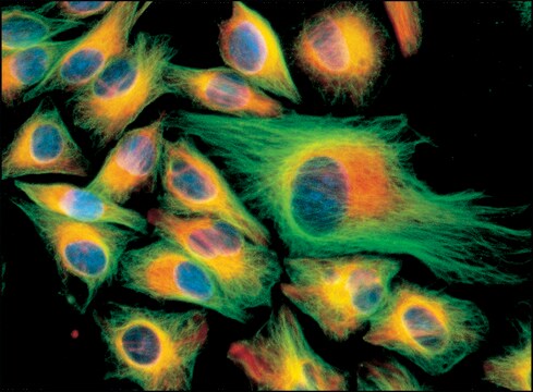
Immunofluorescence (1 paper)
Monoclonal Anti-Cytokeratin, pan antibody produced in mouse has been used in:
  • immunoblotting
  • immunohistochemistry
  • Immunocytochemistry
  • dot blotting

Biochem./physiol. Wirkung

Cytokeratin facilitates the typing of normal, metaplastic, and neoplastic cells and may aid in the discrimination of carcinomas and non-epithelial tumors such as sarcomas, lymphomas, and neural tumors. It is also useful in detecting micrometastases in lymph nodes and other tissues, and for determining the origin of poorly differentiated tumors.

Physikalische Form

Solution in 0.01 M phosphate buffered saline, pH 7.4, containing 15 mM sodium azide.

Haftungsausschluss

Unless otherwise stated in our catalog or other company documentation accompanying the product(s), our products are intended for research use only and are not to be used for any other purpose, which includes but is not limited to, unauthorized commercial uses, in vitro diagnostic uses, ex vivo or in vivo therapeutic uses or any type of consumption or application to humans or animals.

Sie haben nicht das passende Produkt gefunden?  

Probieren Sie unser Produkt-Auswahlhilfe. aus.

Empfehlung

Ähnliches Produkt

Lagerklassenschlüssel

12 - Non Combustible Liquids

WGK

WGK 1

Flammpunkt (°F)

Not applicable

Flammpunkt (°C)

Not applicable


Hier finden Sie alle aktuellen Versionen:

Analysenzertifikate (COA)

Lot/Batch Number

Die passende Version wird nicht angezeigt?

Wenn Sie eine bestimmte Version benötigen, können Sie anhand der Lot- oder Chargennummer nach einem spezifischen Zertifikat suchen.

Besitzen Sie dieses Produkt bereits?

In der Dokumentenbibliothek finden Sie die Dokumentation zu den Produkten, die Sie kürzlich erworben haben.

Die Dokumentenbibliothek aufrufen

Derivation and Characterization of Induced Pluripotent Stem Cells from Equine Fibroblasts
Breton A, et al.
Stem Cells and Development, 22(4), 611-611 (2013)
Pascale Hermant et al.
PloS one, 9(1), e87906-e87906 (2014-02-06)
The type III interferon (IFN) receptor is preferentially expressed by epithelial cells. It is made of two subunits: IFNLR1, which is specific to IFN-lambda (IFN-λ) and IL10RB, which is shared by other cytokine receptors. Human hepatocytes express IFNLR1 and respond
Lina Sun et al.
Scientific reports, 5, 14871-14871 (2015-10-09)
Thymic epithelial cells (TECs) form a 3-dimentional network supporting thymocyte development and maturation. Besides epithelium and thymocytes, heterogeneous fibroblasts are essential components in maintaining thymic microenvironments. However, thymic fibroblast characteristics, development and function remain to be determined. We herein found
A M Di Lullo et al.
Acta otorhinolaryngologica Italica : organo ufficiale della Societa italiana di otorinolaringologia e chirurgia cervico-facciale, 37(3), 207-213 (2016-11-30)
Cystic fibrosis (CF) is an autosomal recessive disease caused by mutations in the cystic fibrosis transmembrane regulator (CFTR) gene. About 2000 mutations have been described so far. We setup an ex vivo model of human nasal epithelial cells (HNECs) to
Nasser K Altorki et al.
Cell reports, 39(1), 110639-110639 (2022-04-07)
To investigate changes in the tumor microenvironment (TME) during lung cancer progression, we interrogate tumors from two chest computed tomography (CT)-defined groups. Pure non-solid (pNS) CT density nodules contain preinvasive/minimally invasive cancers, and solid density nodules contain invasive cancers. Profiling

Unser Team von Wissenschaftlern verfügt über Erfahrung in allen Forschungsbereichen einschließlich Life Science, Materialwissenschaften, chemischer Synthese, Chromatographie, Analytik und vielen mehr..

Setzen Sie sich mit dem technischen Dienst in Verbindung.