
Alle Fotos(1)
Wichtige Dokumente
95349
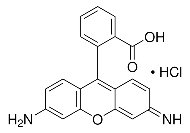
Atto 647N-Amin
BioReagent, suitable for fluorescence
Anmeldenzur Ansicht organisationsspezifischer und vertraglich vereinbarter Preise
Alle Fotos(1)
About This Item
UNSPSC-Code:
12352116
NACRES:
NA.32
Empfohlene Produkte


Produktlinie
BioReagent
Qualitätsniveau
Assay
≥90% (HPLC)
Form
solid
Mol-Gew.
Mw 801 g/mol
Hersteller/Markenname
ATTO-TEC GmbH
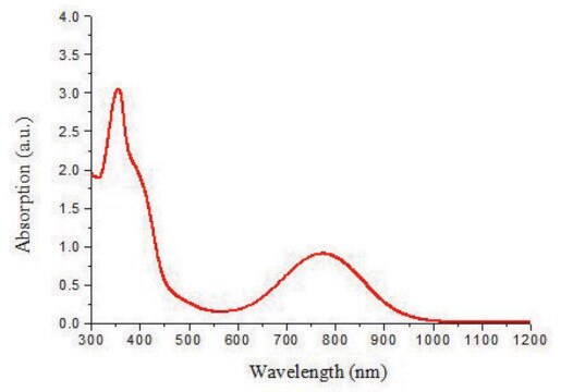
λ
in ethanol (with 0.1% trifluoroacetic acid)
UV-Absorption
λ: 640-646 nm Amax
Eignung
suitable for fluorescence
Lagertemp.
−20°C
Allgemeine Beschreibung
Atto 647N ist ein ausgezeichneter rot emittierender Fluoreszenzfarbstoff mit starker molekularer Absorption (150.000) und Quantenausbeute (0.65) sowie ausreichend großem Stokes-Shift. Atto 647N zeichnet sich durch hohe Photo- und Thermostabilität aus.
Absorption und Fluoreszenz sind unabhängig vom pH-Wert, zumindest in dem am meisten relevanten Bereich von pH 4 bis 11. Das Amin -Derivat kann für Reaktionen mit aktivierten Carboxy-Gruppen, wie NHS-Ester, TFP-Ester usw. eingesetzt werden.
Absorption und Fluoreszenz sind unabhängig vom pH-Wert, zumindest in dem am meisten relevanten Bereich von pH 4 bis 11. Das Amin -Derivat kann für Reaktionen mit aktivierten Carboxy-Gruppen, wie NHS-Ester, TFP-Ester usw. eingesetzt werden.
Lagerklassenschlüssel
11 - Combustible Solids
WGK
WGK 3
Flammpunkt (°F)
Not applicable
Flammpunkt (°C)
Not applicable
Persönliche Schutzausrüstung
Eyeshields, Gloves, type N95 (US)
Hier finden Sie alle aktuellen Versionen:
Besitzen Sie dieses Produkt bereits?
In der Dokumentenbibliothek finden Sie die Dokumentation zu den Produkten, die Sie kürzlich erworben haben.
STED microscopy to monitor agglomeration of silica particles inside A549 cells.
Schubbe, S., et al.
Advanced Engineering Materials, 12, 417-422 (2010)
A novel nanoscopic tool by combining AFM with STED microscopy.
Harke, B., et al.
Optical Nanoscopy, 1, 3-3 (2012)
Volker Westphal et al.
Science (New York, N.Y.), 320(5873), 246-249 (2008-02-23)
We present video-rate (28 frames per second) far-field optical imaging with a focal spot size of 62 nanometers in living cells. Fluorescently labeled synaptic vesicles inside the axons of cultured neurons were recorded with stimulated emission depletion (STED) microscopy in
Marisa L Martin-Fernandez et al.
International journal of molecular sciences, 13(11), 14742-14765 (2012-12-04)
Insights from single-molecule tracking in mammalian cells have the potential to greatly contribute to our understanding of the dynamic behavior of many protein families and networks which are key therapeutic targets of the pharmaceutical industry. This is particularly so at
S E D Webb et al.
Optics express, 16(25), 20258-20265 (2008-12-10)
We combine single molecule fluorescence orientation imaging with single-pair fluorescence resonance energy transfer microscopy, using a total internal reflection microscope. We show how angles and FRET efficiencies can be determined for membrane proteins at the single molecule level and provide
Unser Team von Wissenschaftlern verfügt über Erfahrung in allen Forschungsbereichen einschließlich Life Science, Materialwissenschaften, chemischer Synthese, Chromatographie, Analytik und vielen mehr..
Setzen Sie sich mit dem technischen Dienst in Verbindung.