MABS1249
Anti-SCAP Antibody, clone 9D5
clone 9D5, from mouse
Synonym(e):
Sterol regulatory element-binding protein cleavage-activating protein, SCAP, SREBP cleavage-activating protein
About This Item
Empfohlene Produkte
Biologische Quelle
mouse
Qualitätsniveau
Antikörperform
purified immunoglobulin
Antikörper-Produkttyp
primary antibodies
Klon
9D5, monoclonal
Speziesreaktivität
hamster
Methode(n)
immunocytochemistry: suitable
western blot: suitable
Isotyp
IgG2bκ
NCBI-Hinterlegungsnummer
UniProt-Hinterlegungsnummer
Posttranslationale Modifikation Target
unmodified
Angaben zum Gen
hamster ... Scap(100689048)
Allgemeine Beschreibung
4 lumenal and 5 cytoplasmic regions, having both its N- and C-terminal ends at the cytoplasmic side (a.a. 1-18 & 730-1276). The C-terminal domain of SCAP mediates association with SREBPs, while transmembrane helices 2–6 comprise the sterol-sensing domain that mediates sterol-induced binding of SCAP to Insigs. Mutations within the sterol-sensing region disrupt Insig binding and prevent sterol-mediated ER retention of SCAP-SREBP.
Spezifität
Immunogen
Anwendung
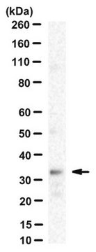
Western Blotting Analysis: A representative lot detected exogenously expressed SCAP co-immunoprecipitated with the exogenously expressed Insig-1 and Insig-2 upon 25-hydroxycholesterol (25-HC) treatment of a SCAP-deficient CHO cell line SRD-13A that had been transfected to co-express SCAP with either myc-tagged Insig-1 or Insig-2 (Lee, P.C., and DeBose-Boyd, R.A. (2010). J. Lipid Res. 51(1):192-201).
Western Blotting Analysis: Representative lots detected similar level of SCAP in the membrane extracts from CHO cells with or without SREBP processing inhibitor 25-hydroxycholesterol (25-HC) treatment regardless whether the cells′ 25-HC-resistant status (Lee, P.C., and DeBose-Boyd, R.A. (2010). J. Lipid Res. 51(1):192-201; Lee, P.C., et al. (2007). J. Lipid Res. 48(9):1944-1954).
Western Blotting Analysis: A representative lot detected the endogenous SCAP immunoprecipitated from CHO cell lysate by a polyclonal SCAP antibody (Sakai, J., et al. (1997). J. Biol. Chem. 272(32):20213-20221).
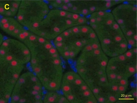
Immunocytochemistry Analysis: A representative lot localized the overexpressed hamster SCAP at ER and nuclear envelope by fluorescent immunocytochemistry staining of 3% paraformaldehyde-fixed, 0.01% saponin-permeabilized CHO stable transfectants (Sakai, J., et al. (1997). J. Biol. Chem. 272(32):20213-20221).
Qualität
Isotyping Analysis: The identity of this monoclonal antibody is confirmed by isotyping test to be IgG2bκ.
Zielbeschreibung
Physikalische Form
Sonstige Hinweise
Sie haben nicht das passende Produkt gefunden?
Probieren Sie unser Produkt-Auswahlhilfe. aus.
Lagerklassenschlüssel
12 - Non Combustible Liquids
WGK
WGK 1
Flammpunkt (°F)
Not applicable
Flammpunkt (°C)
Not applicable
Analysenzertifikate (COA)
Suchen Sie nach Analysenzertifikate (COA), indem Sie die Lot-/Chargennummer des Produkts eingeben. Lot- und Chargennummern sind auf dem Produktetikett hinter den Wörtern ‘Lot’ oder ‘Batch’ (Lot oder Charge) zu finden.
Besitzen Sie dieses Produkt bereits?
In der Dokumentenbibliothek finden Sie die Dokumentation zu den Produkten, die Sie kürzlich erworben haben.
Unser Team von Wissenschaftlern verfügt über Erfahrung in allen Forschungsbereichen einschließlich Life Science, Materialwissenschaften, chemischer Synthese, Chromatographie, Analytik und vielen mehr..
Setzen Sie sich mit dem technischen Dienst in Verbindung.