Direkt zum Inhalt
Merck
Alle Fotos(1)

Wichtige Dokumente

MABE446

Sigma-Aldrich

Anti-Histone H1° Antibody, clone 34

clone 34, from mouse

Synonym(e):

Histone H1.0, Histone H1(0), Histone H1°

Anmeldenzur Ansicht organisationsspezifischer und vertraglich vereinbarter Preise


About This Item

UNSPSC-Code:
12352203
eCl@ss:
32160702
NACRES:
NA.41

Biologische Quelle

mouse

Qualitätsniveau

Antikörperform

purified immunoglobulin

Antikörper-Produkttyp

primary antibodies

Klon

34, monoclonal

Speziesreaktivität

mouse, bovine, Xenopus, human, rat

Speziesreaktivität (Voraussage durch Homologie)

ox (immunogen homology)

Methode(n)

flow cytometry: suitable
immunocytochemistry: suitable
immunohistochemistry: suitable
western blot: suitable

Isotyp

IgG1κ

NCBI-Hinterlegungsnummer

UniProt-Hinterlegungsnummer

Versandbedingung

wet ice

Posttranslationale Modifikation Target

unmodified

Angaben zum Gen

human ... H1F0(3005)

Allgemeine Beschreibung

Histones are highly conserved proteins that serve as the structural scaffold for the organization of nuclear DNA into chromatin. The four core histones, H2A, H2B, H3, and H4, assemble into an octamer (2 molecules of each). Subsequently, 146 base pairs of DNA are wrapped around the octamer, forming a nucleosome. The linker histone, H1, interacts with linker DNA between nucleosomes and functions in the compaction of chromatin into 30nm chromatin fibers and higher order structures.

Spezifität

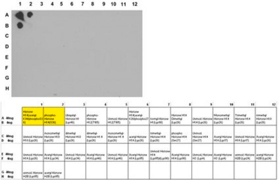
This antibody recognizes Histone H1°.

Immunogen

Recombinant protein corresponding to Ox liver Histone H1°

Anwendung

Research Category
Epigenetik & nukleäre Funktionen
Research Sub Category
Histone
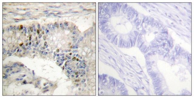
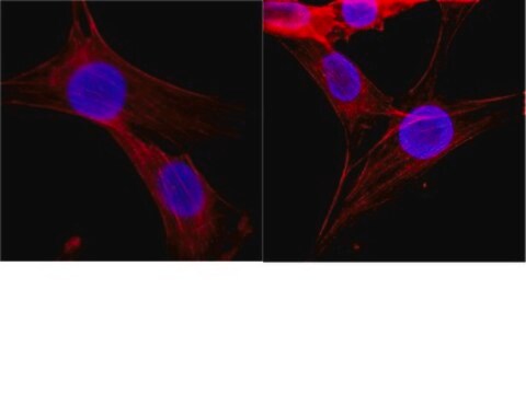
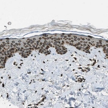
Anti-Histone H1° Antibody, clone 34 is a highly specific mouse monoclonal antibody, that targets Histone H1 & has been tested in western blotting, ICC, IHC & Flow Cytometry.
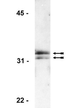
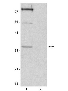
Western Blotting Analysis: A representative lot from an independent laboratory detected Histone H1° in Xenopus embryo tissue lysate (Seigneurin, D., et al. (1995). Int J Dev Biol. 39(4):597-603.; Fu, G., et al. (2003). Biol Reprod. 68(5):1569-1576.).

Immunocytochemistry Analyis: A representative lot from an independent laboratory detected Histone H1° in Xenopus unfertilized eggs and early embryos (Fu, G., et al. (2003). Biol Reprod. 68(5):1569-1576.; Adenot, P. G., et al. (2000). J Cell Sci. 113(Pt 16):2897-2907.).

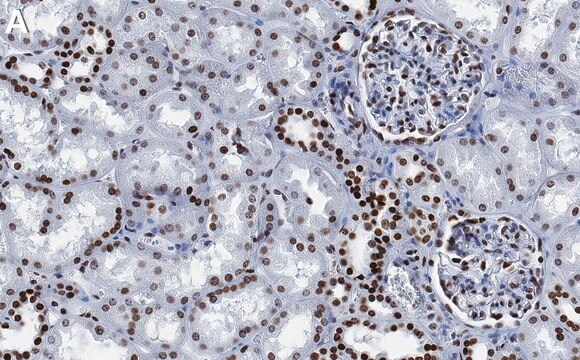
Immunohistochemistry Analysis: A representative lot from an independent laboratory detected Histone H1° in Xenopus embryo tissues (Grunwald, D., et al. (1995). Exp Cell Res. 218(2):586-595.).

Flow Cytometry Analyisis: A representative lot from an independent laboratory detected Histone H1° in FC (Grunwald, D., et al. (1999). Methods Mol Biol. 119:443-454.).

Qualität

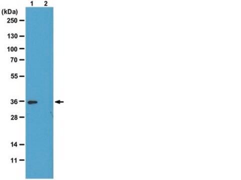
Evaluated by Western Blotting in Jurkat cell lysate.

Western Blotting Analysis: 1 µg/mL of this antibody detected Histone H1° in 10 µg of Jurkat cell lysate.

Zielbeschreibung

~30 kDa observed. Uncharacterized band(s) may be observed in some cell lysates.

Physikalische Form

Protein G Purified
Format: Purified
Purified mouse monoclonal IgG1κ in buffer containing 0.1 M Tris-Glycine (pH 7.4), 150 mM NaCl with 0.05% sodium azide.

Lagerung und Haltbarkeit

Stable for 1 year at 2-8°C from date of receipt.

Hinweis zur Analyse

Control
Jurkat cell lysate

Sonstige Hinweise

Concentration: Please refer to the Certificate of Analysis for the lot-specific concentration.

Haftungsausschluss

Unless otherwise stated in our catalog or other company documentation accompanying the product(s), our products are intended for research use only and are not to be used for any other purpose, which includes but is not limited to, unauthorized commercial uses, in vitro diagnostic uses, ex vivo or in vivo therapeutic uses or any type of consumption or application to humans or animals.

Sie haben nicht das passende Produkt gefunden?  

Probieren Sie unser Produkt-Auswahlhilfe. aus.

Lagerklassenschlüssel

12 - Non Combustible Liquids

WGK

WGK 1

Flammpunkt (°F)

Not applicable

Flammpunkt (°C)

Not applicable


Analysenzertifikate (COA)

Suchen Sie nach Analysenzertifikate (COA), indem Sie die Lot-/Chargennummer des Produkts eingeben. Lot- und Chargennummern sind auf dem Produktetikett hinter den Wörtern ‘Lot’ oder ‘Batch’ (Lot oder Charge) zu finden.

Besitzen Sie dieses Produkt bereits?

In der Dokumentenbibliothek finden Sie die Dokumentation zu den Produkten, die Sie kürzlich erworben haben.

Die Dokumentenbibliothek aufrufen

Germaine Fu et al.
Biology of reproduction, 68(5), 1569-1576 (2003-02-28)
Oocytes and embryos of many species, including mammals, contain a unique linker (H1) histone, termed H1oo in mammals. It is uncertain, however, whether other H1 histones also contribute to the linker histone complement of these cells. Using immunofluorescence and radiolabeling
D Grunwald et al.
Experimental cell research, 218(2), 586-595 (1995-06-01)
It is known that a transition in the linker-histone variants takes place within chromatin during early development of Xenopus laevis; a cleavage-type H1 is replaced by the somatic type. Based on cytofluorimetric analysis of the distribution of the embryo cells
P G Adenot et al.
Journal of cell science, 113 ( Pt 16), 2897-2907 (2000-07-27)
A striking feature of early embryogenesis in a number of organisms is the use of embryonic linker histones or high mobility group proteins in place of somatic histone H1. The transition in chromatin composition towards somatic H1 appears to be
In situ analysis of chromatin proteins during development and cell differentiation using flow cytometry.
D Grunwald et al.
Methods in molecular biology (Clifton, N.J.), 119, 443-454 (2000-05-11)
Developmentally regulated chromatin acetylation and histone H1(0) accumulation.
Seigneurin, D, et al.
International Journal of Developmental Biology, 39, 597-603 (1995)

Unser Team von Wissenschaftlern verfügt über Erfahrung in allen Forschungsbereichen einschließlich Life Science, Materialwissenschaften, chemischer Synthese, Chromatographie, Analytik und vielen mehr..

Setzen Sie sich mit dem technischen Dienst in Verbindung.