Direkt zum Inhalt
Merck

MABE286

Sigma-Aldrich

Anti-Replication Protein A Antibody, clone RPA34-19

clone RPA34-19, from mouse

Synonym(e):

Replication protein A 32 kDa subunit, RP-A p32, Replication factor A protein 2, RF-A protein 2, Replication protein A 34 kDa subunit, RP-A p34

Anmeldenzur Ansicht organisationsspezifischer und vertraglich vereinbarter Preise


About This Item

UNSPSC-Code:
12352203
eCl@ss:
32160702
NACRES:
NA.41

Biologische Quelle

mouse

Qualitätsniveau

Antikörperform

purified antibody

Antikörper-Produkttyp

primary antibodies

Klon

RPA34-19, monoclonal

Speziesreaktivität

human

Methode(n)

immunocytochemistry: suitable
immunohistochemistry: suitable
western blot: suitable

Isotyp

IgG1κ

NCBI-Hinterlegungsnummer

UniProt-Hinterlegungsnummer

Versandbedingung

wet ice

Posttranslationale Modifikation Target

unmodified

Angaben zum Gen

human ... RPA2(6118)

Allgemeine Beschreibung

RPA (RP-A p32) is a heterotrimeric protein complex that binds specifically to single-stranded DNA (ssDNA). It is composed of three subunits: RPA1 (70 kDa), RPA2 (32 kDa), and RPA3 (14 kDa). RPA plays multiple roles in DNA replication. At the onset of DNA replication, RPA is loaded onto chromatin, and is needed for subsequent loading of DNA polymerase and other replication proteins to initiate DNA replication. After replication begins, RPA moves with replication forks, stabilizing ssDNA and assisting in DNA synthesis. In addition to its replication function, RPA is also known to play essential roles in damage repair and recombination. The 32 kDa subunit is phosphorylated by the cdc2 family of kinases when cells enter S-phase; and by ATM, ATR, and DNA-PK proteins in response to DNA damage.

Immunogen

Replication Protein A purified from U293 cells.

Anwendung

Research Category
Epigenetik & nukleäre Funktionen
Research Sub Category
Zellzyklus, DNA-Replikation & -Reparatur
Anti-Replication Protein A Antibody, clone RPA34-19 is a Mouse Monoclonal Antibody for detection of Replication Protein A also known as Replication protein A 32 kDa subunit, RP-A p32 & has been validated in WB, ICC & IHC.
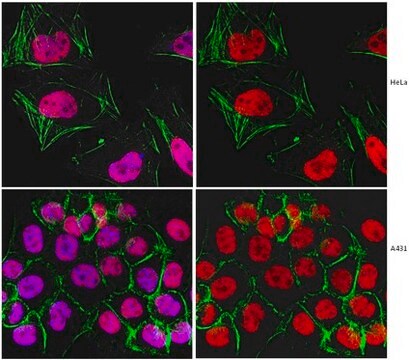
Immunocytochemistry Analysis: A 1:250 dilution from a reprsentative lot detected Replication Protein A in A431 cells.

Immunohistochemistry Analysis: A 1:5 dilution from a representative lot detected Replication Protein A in human placental chorionic villi and in human colorectal adenocarcinoma tissue.

Qualität

Evaluated by Western Blot in HeLa cell lysate.

Western Blot Analysis: A 1:2,000 dilution of this antibody detected Replication Protein A in 10 µg of HeLa cell lysate.

Zielbeschreibung

~34 kDa observed. This protein has 3 isoforms: Isoform 1 (~29 kDa), Isoform 2 (~30 kDa), and Isoform 3 (~39 kDa).

Physikalische Form

Protein G Purified
Format: Purified
Purified mouse monoclonal IgG1κ in buffer containing 0.1 M Tris-Glycine (pH 7.4), 150 mM NaCl with 0.05% sodium azide.

Lagerung und Haltbarkeit

Stable for 1 year at 2-8°C from date of receipt.

Hinweis zur Analyse

Control
HeLa cell lysate

Haftungsausschluss

Unless otherwise stated in our catalog or other company documentation accompanying the product(s), our products are intended for research use only and are not to be used for any other purpose, which includes but is not limited to, unauthorized commercial uses, in vitro diagnostic uses, ex vivo or in vivo therapeutic uses or any type of consumption or application to humans or animals.

Sie haben nicht das passende Produkt gefunden?  

Probieren Sie unser Produkt-Auswahlhilfe. aus.

Lagerklassenschlüssel

12 - Non Combustible Liquids

WGK

WGK 1

Flammpunkt (°F)

Not applicable

Flammpunkt (°C)

Not applicable


Analysenzertifikate (COA)

Suchen Sie nach Analysenzertifikate (COA), indem Sie die Lot-/Chargennummer des Produkts eingeben. Lot- und Chargennummern sind auf dem Produktetikett hinter den Wörtern ‘Lot’ oder ‘Batch’ (Lot oder Charge) zu finden.

Besitzen Sie dieses Produkt bereits?

In der Dokumentenbibliothek finden Sie die Dokumentation zu den Produkten, die Sie kürzlich erworben haben.

Die Dokumentenbibliothek aufrufen

Motohiro Yamauchi et al.
DNA repair, 7(3), 405-417 (2008-02-06)
Several DNA damage checkpoint factors form nuclear foci in response to ionizing radiation (IR). Although the number of the initial foci decreases concomitantly with DNA double-strand break repair, some fraction of foci persists. To date, the physiological role of the
Janna Luessing et al.
iScience, 25(7), 104536-104536 (2022-06-28)
Abscission, the final stage of cytokinesis, occurs when the cytoplasmic canal connecting two emerging daughter cells is severed either side of a large proteinaceous structure, the midbody. Here, we expand the functions of ATR to include a cell-cycle-specific role in

Unser Team von Wissenschaftlern verfügt über Erfahrung in allen Forschungsbereichen einschließlich Life Science, Materialwissenschaften, chemischer Synthese, Chromatographie, Analytik und vielen mehr..

Setzen Sie sich mit dem technischen Dienst in Verbindung.