MAB5278
Anti-Tryptophan Hydroxylase/Tyrosine Hydroxylase/Phenylalanine Hydroxylase Antibody, clone PH8
clone PH8, Chemicon®, from mouse
About This Item
Empfohlene Produkte
Biologische Quelle
mouse
Qualitätsniveau
Antikörperform
purified immunoglobulin
Antikörper-Produkttyp
primary antibodies
Klon
PH8, monoclonal
Speziesreaktivität
human
Hersteller/Markenname
Chemicon®
Methode(n)
immunohistochemistry: suitable
immunoprecipitation (IP): suitable
western blot: suitable
Isotyp
IgG1
NCBI-Hinterlegungsnummer
Versandbedingung
wet ice
Posttranslationale Modifikation Target
unmodified
Angaben zum Gen
human ... TPH1(7166)
Allgemeine Beschreibung
Anwendung
PROTOCOLS
Immunohistochemistry
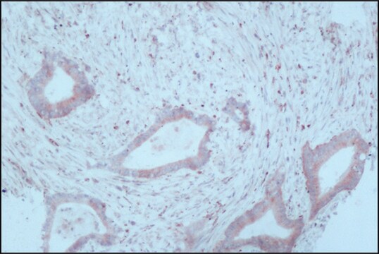
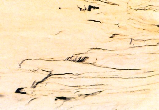
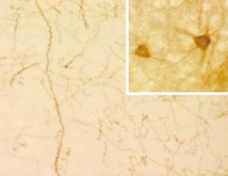
The PH8 Antibody can be used for the immunohistochemical detection of dopaminergic and serotonergic neurons in human and rat brain stem tissue.
1. Tissue should be formalin fixed and stored in formalin prior to use for a minimum of five days.
2. Cryoprotect tissue using 30% sucrose in 0.1M Tris pH 7.4 buffer for 24-72 hours.
3. Cut using a sledge microtome to 50 μm thickness.
4. Wash the tissue samples using Tris buffer prior to commencing the staining procedure.
5. Treat the tissue samples for 3 x 15 minutes in 50% alcohol.
6. Treat the tissue samples for 20 minutes in 50% alcohol and 3% H2O2.
7. Treat tissue for 20 minutes with 10% normal horse serum in Tris buffer. This acts to block endogeneous H2O2 staining.
8. Dilute the PH8 Antibody in Tris buffer, add to the tissue samples and incubate for 1-3 days. Recommended dilutions are 1:2,000-1:10,000. At high antibody concentration all monoaminergic neurons are stained with no distinction between serotonegic and catecholominergic cells. By diluting the antibody concentrate, cell types are distinguishable due to variation in staining intensity. At lower concentrations (1:5,000-1:10,000 dilution) only serotonergic cells will stain.
9. Wash tissue (3 x 15 minutes), then add a biotinylated anti-mouse secondary antibody and incubate on an orbital shaker at room temperature (RT) for 1 hour.
10. Wash tissue (3 x 15 minutes) and incubate on an orbital shaker at RT for 1 hour with the tertiary complex (ELITE KIT, Vector, USA).
11. Wash tissue (3 x 15 minutes) and incubate on an orbital shaker at RT for 10 minutes with Tris buffered diamino-benzidine substrate. Add 0.1% H2O2 and incubate on an orbital shaker at RT for a further 5 minutes.
12. Mount tissue onto gelatinised slides and allow to dry prior to microscopy.
Physikalische Form
Lagerung und Haltbarkeit
Rechtliche Hinweise
Sie haben nicht das passende Produkt gefunden?
Probieren Sie unser Produkt-Auswahlhilfe. aus.
Lagerklassenschlüssel
12 - Non Combustible Liquids
WGK
WGK 2
Flammpunkt (°F)
Not applicable
Flammpunkt (°C)
Not applicable
Analysenzertifikate (COA)
Suchen Sie nach Analysenzertifikate (COA), indem Sie die Lot-/Chargennummer des Produkts eingeben. Lot- und Chargennummern sind auf dem Produktetikett hinter den Wörtern ‘Lot’ oder ‘Batch’ (Lot oder Charge) zu finden.
Besitzen Sie dieses Produkt bereits?
In der Dokumentenbibliothek finden Sie die Dokumentation zu den Produkten, die Sie kürzlich erworben haben.
Unser Team von Wissenschaftlern verfügt über Erfahrung in allen Forschungsbereichen einschließlich Life Science, Materialwissenschaften, chemischer Synthese, Chromatographie, Analytik und vielen mehr..
Setzen Sie sich mit dem technischen Dienst in Verbindung.
