Accéder au contenu
MilliporeSigma
Toutes les photos(1)

Key Documents

M4278

Sigma-Aldrich

Monoclonal Anti-MAP1 antibody produced in mouse

clone HM-1, ascites fluid

Synonyme(s) :

Anti-MAP1a

Se connecterpour consulter vos tarifs contractuels et ceux de votre entreprise/organisme


About This Item

Numéro MDL:
Code UNSPSC :
12352203
Nomenclature NACRES :
NA.41

Source biologique

mouse

Niveau de qualité

Conjugué

unconjugated

Forme d'anticorps

ascites fluid

Type de produit anticorps

primary antibodies

Clone

HM-1, monoclonal

Contient

15 mM sodium azide

Espèces réactives

mouse, rat

Technique(s)

immunohistochemistry (frozen sections): suitable
microarray: suitable
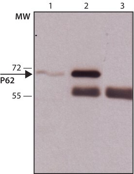
western blot: 1:500 using a fresh total rat brain extract or an enriched microtubule protein preparation

Isotype

IgG1

Numéro d'accès UniProt

Conditions d'expédition

dry ice

Température de stockage

−20°C

Modification post-traductionnelle de la cible

unmodified

Informations sur le gène

mouse ... Mtap1a(17754)
rat ... Map1a(25152)

Description générale

Monoclonal Anti-MAP1 (mouse IgG1 isotype) is derived from the HM-1 hybridoma produced by the fusion of mouse myeloma cells and splenocytes from BALB/c mice immunized with a rat brain MAPs preparation. MAP1 is one of the major neuronal MAPs as well as being the largest (350 kD). MAP1 is more generally distributed, being found in both dendrites and axons of neurons and in glial cells in brain, in chromatophores and on both interphase and mitotic microtubules in various tissue culture cells.

Spécificité

The antibody does not cross-react with other MAPs or tubulin. By immunohistochemical staining of brain tissue, the antibody shows selective labeling of neurons with stronger staining of axons than dendrites.

Immunogène

rat brain microtubule-associated proteins (MAPs)

Application

Monoclonal Anti-MAP1 has been used in :
  • immunoblotting
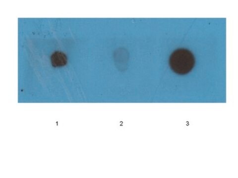
  • dot blot
  • immunocytochemistry
  • immunohistochemistry

Actions biochimiques/physiologiques

Microtubules are the ubiquitous cytoskeletal structural components that are involved in intracellular transport. They are composed of tubulin and microtubule-associated proteins (MAPs). MAPs are known to mediate the binding of membranous organelles, actin filaments and intermediate filaments to microtubules. Therefore, it might be important for cellular processes such as mitosis and organelle transport, and for determining the dynamic properties of the cytoskeleton.

Clause de non-responsabilité

Unless otherwise stated in our catalog or other company documentation accompanying the product(s), our products are intended for research use only and are not to be used for any other purpose, which includes but is not limited to, unauthorized commercial uses, in vitro diagnostic uses, ex vivo or in vivo therapeutic uses or any type of consumption or application to humans or animals.

Vous ne trouvez pas le bon produit ?  

Essayez notre Outil de sélection de produits.

Code de la classe de stockage

10 - Combustible liquids

Classe de danger pour l'eau (WGK)

WGK 1


Certificats d'analyse (COA)

Recherchez un Certificats d'analyse (COA) en saisissant le numéro de lot du produit. Les numéros de lot figurent sur l'étiquette du produit après les mots "Lot" ou "Batch".

Déjà en possession de ce produit ?

Retrouvez la documentation relative aux produits que vous avez récemment achetés dans la Bibliothèque de documents.

Consulter la Bibliothèque de documents

The calcium-sensitive large-conductance potassium channel (BK/MAXI K) is present in the inner mitochondrial membrane of rat brain
Douglas RM, et al.
Neuroscience, 139(4), 1249-1261 (2006)
Effect of intranasal administration of semliki forest virus recombinant particles expressing reporter and cytokine genes on the progression of experimental autoimmune encephalomyelitis
Jerusalmi A, et al.
Molecular Therapy, 8(6), 886-894 (2003)
Cytoskeletons in the Closet-Subversion in Alphaherpesvirus Infections
Denes C, et al.
Viruses, 10(2), 79-79 (2018)
Drosophila Futsch/22C10 is a MAP1B-like protein required for dendritic and axonal development
Hummel T, et al.
Neuron, 26(2), 357-370 (2000)
Alejandra Bosco et al.
The Journal of comparative neurology, 519(4), 599-620 (2011-01-20)
Changes in microglial cell activation and distribution are associated with neuronal decline in the central nervous system (CNS), particularly under pathological conditions. Activated microglia converge on the initial site of axonal degeneration in human glaucoma, yet their part in its

Notre équipe de scientifiques dispose d'une expérience dans tous les secteurs de la recherche, notamment en sciences de la vie, science des matériaux, synthèse chimique, chromatographie, analyse et dans de nombreux autres domaines..

Contacter notre Service technique