Accéder au contenu
MilliporeSigma
Toutes les photos(1)

Principaux documents

C7923

Sigma-Aldrich

Monoclonal Anti-CD44 antibody produced in mouse

clone A3D8, purified immunoglobulin, buffered aqueous solution

Synonyme(s) :

Anti-CDW44, Anti-CSPG8, Anti-ECMR-III, Anti-HCELL, Anti-HUTCH-I, Anti-IN, Anti-LHR, Anti-MC56, Anti-MDU2, Anti-MDU3, Anti-MIC4, Anti-Pgp1

Se connecterpour consulter vos tarifs contractuels et ceux de votre entreprise/organisme


About This Item

Numéro MDL:
Code UNSPSC :
12352203
Nomenclature NACRES :
NA.41
Conjugué:
unconjugated
application:
FACS
Clone:
A3D8, monoclonal
Espèces réactives:
human
citations:
16
Technique(s):
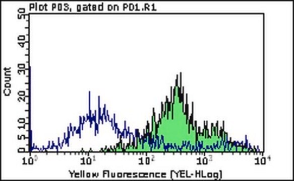
flow cytometry: 5 μL using 1 × 10⁶ cells

Source biologique

mouse

Conjugué

unconjugated

Forme d'anticorps

purified immunoglobulin

Type de produit anticorps

primary antibodies

Clone

A3D8, monoclonal

Forme

buffered aqueous solution

Poids mol.

antigen 80-95 kDa

Espèces réactives

human

Technique(s)

flow cytometry: 5 μL using 1 × 10⁶ cells

Isotype

IgG1

Numéro d'accès UniProt

Conditions d'expédition

wet ice

Température de stockage

2-8°C

Modification post-traductionnelle de la cible

unmodified

Informations sur le gène

human ... CD44(960)

Description générale

CD44 is a glycosylated cartilage-linked protein. The protein exists in a variety of forms with different molecular structures, ranging from 85 kDa to 250 kDa. It is a multistructural and multifunctional cell surface molecule and involved in various biological processes such as cell proliferation, cell differentiation, cell migration, angiogenesis, presentation of cytokines, chemokines, and growth factors to the corresponding receptors, and docking of proteases at the cell membrane, as well as in signaling for cell survival. Although all the activities are related to normal cell, but it has also some pathologic activities of cancer cells.

Spécificité

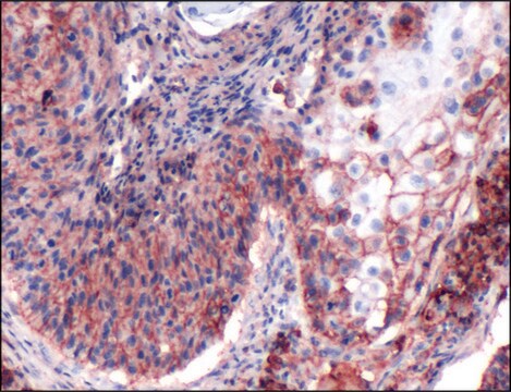
Recognizes the CD44 (80-95 kDa) human cell surface glycoprotein on B and T lymphocytes, monocytes, granulocytes and red blood cells. The epitope recognized by this antibody is sensitive to bromelain and other proteolytic enzymes. It is detectable in routine formalin-fixed, paraffin-embedded tissue.
5th Workshop: code no. BP406, S320

Immunogène

circulating malignant human Sezary T cells.

Application

Applications in which this antibody has been used successfully, and the associated peer-reviewed papers, are given below.
Flow cytometry/Cell sorting (1 paper)
Monoclonal Anti-CD44 antibody was used as a hyaluronic acid receptor for indirect immunoperoxidase technique in a study to evaluate the relationship of lymphoid cells and macrophages with vasculature and stromal components.
Anti CD44 monoclonal antibody was used for:
  • immunostaining of nonpapillary carcinoma fine-needle aspiration (FNA) or surgical excision specimen.
  • flow cytometry analysis to assess breast CSCs in human MCF-7/Dox breast cancer cells.
  • staining of CD44 in immunohistochemistry analysis.
  • incubation of osteoclasts (OCs) in a study to analyze the molecular composition and F-actin organization of the OC podosome belts.
  • It is also suitable for western blot at a dilution of 1:500-1:2,000

Actions biochimiques/physiologiques

The CD44 antigen is expressed on a variety of cell types including peripheral blood leukocytes (B and T lymphocytes, monocytes, granuloctes) and red cells. It is also weakly expressed on platelets. The antibody is also reactive with bone marrow nucleated cells, medullary thymocytes, liver Kupffer cells, fibroblasts, corneal cells, epidermal keratinocytes, synovial cells, a subset of pancreatic acinar cells and brain cells. The epitope recognized by this clone is sensitive to formalin fixation and paraffin embedding.

Description de la cible

The CD44 antigen has been studied under the following names: Pgp-1, Hermes antigen, ECM-III, H-CAM and HUTCH-1. It functions as a homing receptor. CD44 antigen is the cellular receptor for hyaluronic acid. The antibody is also reactive with bone marrow nucleated cells, medullary thymocytes, liver Kupffer cells, fibroblasts, corneal cells, epidermal keratinocytes, synovial cells, a subset of pancreatic acinar cells and brain cells.

Forme physique

Solution in 0.01 M phosphate buffered saline, pH 7.4, containing 1% bovine serum albumin and 15 mM sodium azide.

Stockage et stabilité

Store at 2-8°C for up to one month. For extended storage, the solution may be frozen in working aliquots. Repeated freezing and thawing, or storage in "frost-free" freezers, is not recommended. If slight turbidity occurs upon prolonged storage, clarify the solution by centrifugation before use.

Clause de non-responsabilité

Unless otherwise stated in our catalog or other company documentation accompanying the product(s), our products are intended for research use only and are not to be used for any other purpose, which includes but is not limited to, unauthorized commercial uses, in vitro diagnostic uses, ex vivo or in vivo therapeutic uses or any type of consumption or application to humans or animals.

Vous ne trouvez pas le bon produit ?  

Essayez notre Outil de sélection de produits.

Code de la classe de stockage

10 - Combustible liquids

Classe de danger pour l'eau (WGK)

nwg

Point d'éclair (°F)

Not applicable

Point d'éclair (°C)

Not applicable


Faites votre choix parmi les versions les plus récentes :

Certificats d'analyse (COA)

Lot/Batch Number

Vous ne trouvez pas la bonne version ?

Si vous avez besoin d'une version particulière, vous pouvez rechercher un certificat spécifique par le numéro de lot.

Déjà en possession de ce produit ?

Retrouvez la documentation relative aux produits que vous avez récemment achetés dans la Bibliothèque de documents.

Consulter la Bibliothèque de documents

Goar Mosoyan et al.
PloS one, 8(1), e55145-e55145 (2013-02-02)
Breast cancer cell lines are widely used tools to investigate breast cancer biology and to develop new therapies. Breast cancer tissue contains molecularly heterogeneous cell populations. Thus, it is important to understand which cell lines best represent the primary tumor
Vineet Gupta et al.
Methods in molecular biology (Clifton, N.J.), 716, 179-191 (2011-02-15)
Flow cytometry can sensitively detect and efficiently sort cells based on fluorescent signals integrated into cellular markers of proteins or DNA. It has been broadly applied to assess cell division, apoptosis and to isolate cells including stem cells. As the
B F Haynes et al.
Immunology today, 10(12), 423-428 (1989-12-01)
The study of cell surface molecules that are involved in interactions between immune and non-hematopoietic cells in various microenvironments is currently an area of great interest. One molecule that appears to be involved in multiple steps of normal immune cell
D C Chhieng et al.
Cancer, 81(3), 157-162 (1997-06-25)
Although nuclear grooves and inclusions are considered to be characteristic cytologic features of thyroid papillary carcinoma, a variety of other thyroid lesions may on occasion display these features in fine-needle aspiration specimens. The authors evaluated the immunocytochemical staining of 16
Abraham Zepeda-Moreno et al.
Cell adhesion & migration, 5(3), 215-219 (2011-02-23)
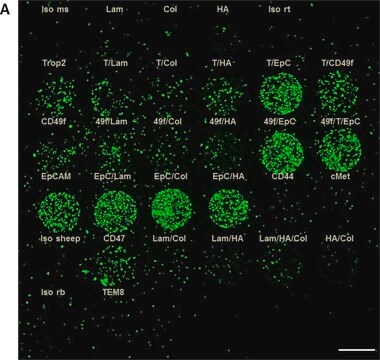
Cell adhesion is an important part of many complex biological processes. It plays crucial roles in cancer, development, and maintenance of stem cell compartment. The measurement of adhesion under experimental conditions might provide important information for cell biology. There are

Articles

Cancer stem cell media, spheroid plates and cancer stem cell markers to culture and characterize CSC populations.

Cancer stem cell media, spheroid plates and cancer stem cell markers to culture and characterize CSC populations.

Notre équipe de scientifiques dispose d'une expérience dans tous les secteurs de la recherche, notamment en sciences de la vie, science des matériaux, synthèse chimique, chromatographie, analyse et dans de nombreux autres domaines..

Contacter notre Service technique