Accéder au contenu
MilliporeSigma
Toutes les photos(1)

Key Documents

NE1022

Sigma-Aldrich

PhosphoDetect Anti-Neurofilament H Mouse mAb (SMI-31)

liquid, clone SMI-31, Calbiochem®

Synonyme(s) :

Anti-neurofilament antibody

Se connecterpour consulter vos tarifs contractuels et ceux de votre entreprise/organisme


About This Item

Code UNSPSC :
12352203
Nomenclature NACRES :
NA.41

Source biologique

mouse

Niveau de qualité

Forme d'anticorps

purified antibody

Type de produit anticorps

primary antibodies

Clone

SMI-31, monoclonal

Forme

liquid

Contient

≤0.1% sodium azide as preservative

Espèces réactives

chicken, Xenopus

Réactivité de l'espèce (prédite par homologie)

mammals

Fabricant/nom de marque

Calbiochem®

Conditions de stockage

OK to freeze
avoid repeated freeze/thaw cycles

Isotype

IgG1

Conditions d'expédition

wet ice

Température de stockage

2-8°C

Modification post-traductionnelle de la cible

unmodified

Informations sur le gène

human ... NEFH(4744)

Description générale

Mouse monoclonal antibody supplied as purified antibody. Recognizes the ~180-200 kDa phosphorylated neurofilament H protein.
Recognizes the ~180-200 kDa phosphorylated neurofilament H protein in rat central nervous system (CNS) cytoskeletal preparations. Also weakly recognizes phosphorylated NF-M protein.
This PhosphoDetect Anti-Neurofilament H Mouse mAb (SMI-31) is validated for use in ELISA, Frozen Sections, Immunoblotting, ICC, Paraffin Sections, IP for the detection of Neurofilament H.

Immunogène

homogenized, hypothalami from Fischer 344 rat brain

Application


ELISA (1:1000)
Frozen Sections (1:1000, see comments)
Immunoblotting (1:1000)
Immunocytochemistry (1:1000, see comments)
Paraffin Sections (1:1000, heat pre-treatment required, see comments)
Immunoprecipitation (see comments)

Avertissement

Toxicity: Standard Handling (A)

Forme physique

In PBS and Thimerosal

Autres remarques

Raina, A.K., et al. 1999. Neuroreport10, 1355.
Yang, C.C., et al. 1998. Brain121, 1089.
Giasson, B.I and Mushynski, W.E. 1996. J. Biol. Chem.271, 30404.
Mirabella, M., et al. 1996. J. Neuropath. Exp. Neurol.55, 774.
Xiao, J. and Monteiro, M.J. 1994. J. Neurosci.14, 1820.
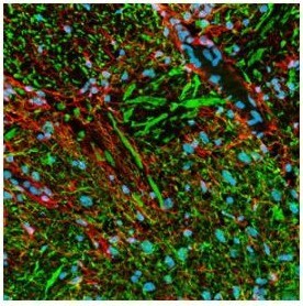
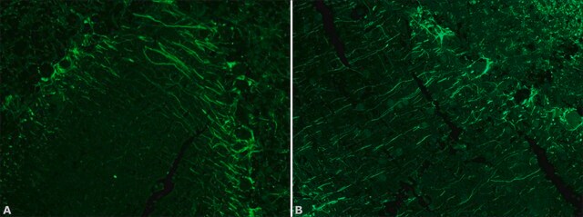
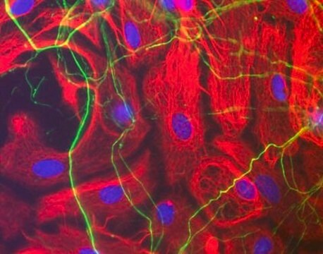
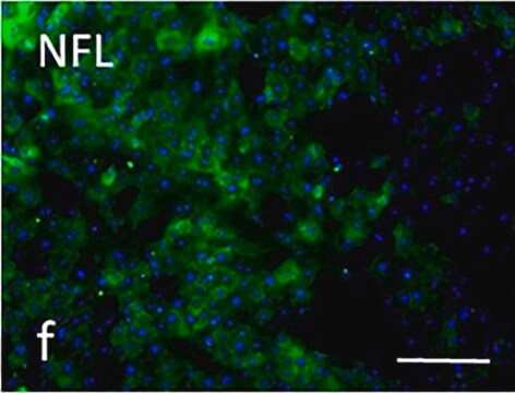
Strongly recognizes phosphorylated neurofilament H and, to a lesser extent, phosphorylated neurofilament M. By immunocytochemistry this antibody broadly stains thick and thin axons and some dendrites, such as basket cell dendrites, but not Purkinje cell dendrites. Does not generally stain nerve cell bodies or other cells and tissues except peripheral axons. May also stain neuronal cell bodies in pathological conditions. Aberrant phosphorylation of neurofilament H in cell bodies can be demonstrated in neuronal cell cultures following treatment with agents that induce stress-activated protein kinase. This antibody is reported to co-immunoprecipitate neurofilament-associated kinase (NAK 115) via interaction of the antibody with the tail domain of neurofilament H. Phosphatase treatment of tissue sections or immunoblotting samples abolishes antibody reactivity. Reactivity is unaffected by trypsin treatment of samples. Tissues and cultured cells can be fixed with a variety of paraformaldehyde- or formaldehyde-containing fixatives, including Bouin′s fixative. Post-fixation in cold methanol or methanol/hydrogen peroxide facilitates access of the antibody to the epitope in frozen sections or thick tissue sections fixed in 4% paraformaldehyde and in cultured cells. For staining formalin-fixed, paraffin sections it is recommended that the de-paraffinized tissue be autoclaved in dH2O for 10 min or boiled in Tris-buffered saline, pH 9.0, in a microwave for 15 min to expose the epitope. Antibody should be titrated for optimal results in individual systems.

Informations légales

CALBIOCHEM is a registered trademark of Merck KGaA, Darmstadt, Germany

Vous ne trouvez pas le bon produit ?  

Essayez notre Outil de sélection de produits.

Code de la classe de stockage

12 - Non Combustible Liquids

Classe de danger pour l'eau (WGK)

nwg

Point d'éclair (°F)

Not applicable

Point d'éclair (°C)

Not applicable


Certificats d'analyse (COA)

Recherchez un Certificats d'analyse (COA) en saisissant le numéro de lot du produit. Les numéros de lot figurent sur l'étiquette du produit après les mots "Lot" ou "Batch".

Déjà en possession de ce produit ?

Retrouvez la documentation relative aux produits que vous avez récemment achetés dans la Bibliothèque de documents.

Consulter la Bibliothèque de documents

Masamitsu Shimazawa et al.
Journal of neuroscience research, 90(10), 1960-1969 (2012-06-08)
We evaluated time-dependent optic nerve degeneration and the role of endoplasmic reticulum (ER) stress in this process following retinal ganglion cell death in mice. Retinal damage was induced by intravitreal injection of N-methyl-D-aspartate (NMDA). Neurofilament heavy (NFH)- and phosphorylated NFH
Seungyong Lee et al.
Nature communications, 12(1), 4939-4939 (2021-08-18)
Pain is a central feature of soft tissue trauma, which under certain contexts, results in aberrant osteochondral differentiation of tissue-specific stem cells. Here, the role of sensory nerve fibers in this abnormal cell fate decision is investigated using a severe
Justin C Burrell et al.
Journal of biomedical materials research. Part A, 109(7), 1183-1195 (2020-09-29)
Promising biomaterials should be tested in appropriate large animal models that recapitulate human inflammatory and regenerative responses. Previous studies have shown tyrosine-derived polycarbonates (TyrPC) are versatile biomaterials with a wide range of applications across multiple disciplines. The library of TyrPC
Chongbo Zhong et al.
Frontiers in neural circuits, 16, 978837-978837 (2022-10-11)
Modulation of the release of glutamate by activation of presynaptic nicotinic acetylcholine receptors (nAChRs) is one of the most prevalent mechanism of nicotinic facilitation of glutamatergic transmission in cortico-limbic circuits. By imaging gene chimeric co-cultures from mouse, we examined the
Michael J Whitehead et al.
Scientific reports, 8(1), 5219-5219 (2018-03-28)
Axon degeneration underlies many nervous system diseases; therefore understanding the regulatory signalling pathways is fundamental to identifying potential therapeutics. Previously, we demonstrated heparan sulphates (HS) as a potentially new target for promoting CNS repair. HS modulate cell signalling by both

Notre équipe de scientifiques dispose d'une expérience dans tous les secteurs de la recherche, notamment en sciences de la vie, science des matériaux, synthèse chimique, chromatographie, analyse et dans de nombreux autres domaines..

Contacter notre Service technique