Accéder au contenu
MilliporeSigma
Toutes les photos(3)

Principaux documents

MAB1273B

Sigma-Aldrich

Anti-Mitochondria Antibody, clone 113-1, Biotin Conjugate

clone 113-1, from mouse, biotin conjugate

Synonyme(s) :

Human Mitochondria

Se connecterpour consulter vos tarifs contractuels et ceux de votre entreprise/organisme


About This Item

Code UNSPSC :
12352203
eCl@ss :
32160702
Nomenclature NACRES :
NA.41

Source biologique

mouse

Niveau de qualité

Conjugué

biotin conjugate

Forme d'anticorps

purified immunoglobulin

Type de produit anticorps

primary antibodies

Clone

113-1, monoclonal

Espèces réactives

human (mitochondria protein)

Ne doit pas réagir avec

rat (mitochondria protein), mouse (mitochondria protein)

Technique(s)

immunocytochemistry: suitable
immunohistochemistry: suitable

Isotype

IgG1

Conditions d'expédition

wet ice

Modification post-traductionnelle de la cible

unmodified

Description générale

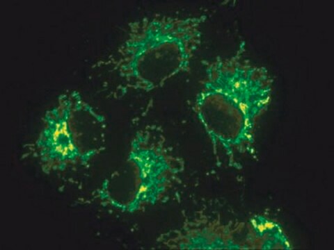
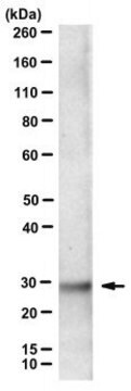
This antibody recognizes a 60 kDa nonglycosylated protein component of mitochondria found in human cells. It can be used to stain mitochondria in cell, tissue or biochemical preparations and can be used as a marker of the mitochondria in subcellular fractions. MAB1273B recognizes a 65 kD protein by immunoprecipitation and produces the stringy, "spaghetti-like", staining patten in the cytoplasm of human cells.

Spécificité

This antibody recognizes human. Antibody is not reactive with mouse or rodent.

Immunogène

Surface of intact human mitochondria

Application

Detect Mitochondrial Marker using this mouse monoclonal antibody, Anti-Mitochondria Antibody, clone 113-1, Biotin Conjugate validated for use in ICC & IHC.
Evaluated by Immunocytochemistry in primary mouse embryonic fibroblasts (PMEFs).
Immunocytochemsitry Analysis: A 1:50 dilution of this antibody did not detect mitrochondria in primary mouse embryonic fibroblasts (PMEFs). Evaluated by Immunohistochemistry in human kidney tissue.
Immunocytochemsitry Analysis: A 1:500 dilution of this antibody detected mitrochondria in human kidney tissue.
Research Category
Stem Cell Research

Cell Structure
Research Sub Category
Developmental Neuroscience

Organelle & Cell Markers

Qualité

Evaluated by Immunocytochemistry in human adipose mesenchymal stem cells (SCC038).
Immunocytochemsitry Analysis: A 1:50 dilution of this antibody detected mitrochondria in human adipose mesenchymal stem cells (SCC038).

Description de la cible

60 kDa calculated

Forme physique

Protein G Purified
Purified mouse monoclonal IgG1 conjugated to Biotin in PBS with 0.1% sodium azide and 15mg/ml BSA.

Stockage et stabilité

Maintain refrigerated at 2-8ºC protected from light in undiluted aliquots for up to 6 months from date of receipt.

Remarque sur l'analyse

Control
Human adipose mesenchymal stem cells (SCC038).

Autres remarques

Concentration: Please refer to the Certificate of Analysis for the lot-specific concentration.

Clause de non-responsabilité

Unless otherwise stated in our catalog or other company documentation accompanying the product(s), our products are intended for research use only and are not to be used for any other purpose, which includes but is not limited to, unauthorized commercial uses, in vitro diagnostic uses, ex vivo or in vivo therapeutic uses or any type of consumption or application to humans or animals.

Vous ne trouvez pas le bon produit ?  

Essayez notre Outil de sélection de produits.

Code de la classe de stockage

12 - Non Combustible Liquids

Classe de danger pour l'eau (WGK)

WGK 2

Point d'éclair (°F)

Not applicable

Point d'éclair (°C)

Not applicable


Certificats d'analyse (COA)

Recherchez un Certificats d'analyse (COA) en saisissant le numéro de lot du produit. Les numéros de lot figurent sur l'étiquette du produit après les mots "Lot" ou "Batch".

Déjà en possession de ce produit ?

Retrouvez la documentation relative aux produits que vous avez récemment achetés dans la Bibliothèque de documents.

Consulter la Bibliothèque de documents

Emily Pulford et al.
Pathology, 48(7), 650-659 (2016-12-14)
Vasculogenic mimicry, the process in which cancer cells form angiomatoid structures independent of or in addition to host angiogenesis has been recorded in several otherwise non-endothelial malignant neoplasms. This study describes evidence of routine vascular mimicry by human mesothelioma cell
Kai Kang et al.
Biomaterials, 230, 119574-119574 (2019-11-26)
Engineered heart tissues (EHTs) are regarded as being the most promising alternative to synthetic materials, and autologous mesenchymal stem cells (MSCs) are widely used as seeding cells. However, few studies have evaluated the feasibility of using MSCs from patients with

Notre équipe de scientifiques dispose d'une expérience dans tous les secteurs de la recherche, notamment en sciences de la vie, science des matériaux, synthèse chimique, chromatographie, analyse et dans de nombreux autres domaines..

Contacter notre Service technique