ECM506
QCM Chemotaxis Cell Migration Assay, 24-well (5 µm), colorimetric
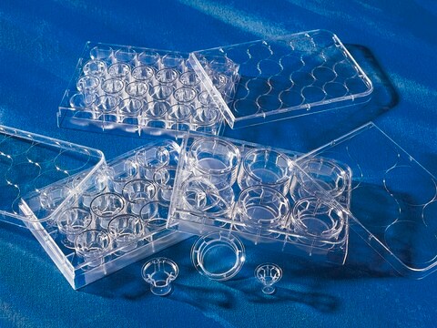
This 3 um QCM Chemotaxis Assay 24-well plate -colorimetric is performed in a Migration Chamber, based on the Boyden chamber principle.
Synonyme(s) :
Cell migration assay, Chemotaxis assay
Se connecterpour consulter vos tarifs contractuels et ceux de votre entreprise/organisme
About This Item
Code UNSPSC :
12352207
eCl@ss :
32161000
Nomenclature NACRES :
NA.84
Produits recommandés
Niveau de qualité
Réactivité de l'espèce (prédite par homologie)
all
Fabricant/nom de marque
Chemicon®
QCM
Technique(s)
activity assay: suitable
cell based assay: suitable
Méthode de détection
colorimetric
Conditions d'expédition
wet ice
Description générale
Also available: Cell Comb Scratch Assay! Get biochemical data from a scratch assay!Click Here
Introduction
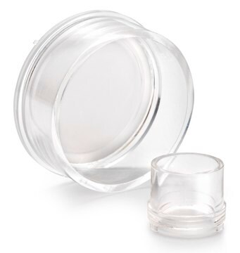
Cell migration is a fundamental function of normal cellular processes, including embryonic development, angiogenesis, wound healing, immune response, and inflammation (1, 2). Most cells are sized from 30 to 50 µm can migrate through 3 to 10 µm pore. Microporous membrane inserts are widely used for cell migration and invasion assays. The most widely accepted of which is the Boyden Chamber assay. The Boyden Chamber system uses a hollow plastic chamber, sealed at one end with a porous membrane. This chamber is suspended over a larger well which may contain medium and/or chemoattractants. Cells are placed inside the Chamber and allowed to migrate through the pores, to the other side of the membrane. Migratory cells are then stained and counted. Most migration assays utilize an 8 µm pore size, as this is appropriate for most cell types, e.g. epithelial and fibroblast cells. The Millipore 5 µm QCM Chemotaxis Assay 24-well- colorimetric utilizes a 5 µm pore size, which is appropriate for a subset of fibroblast cells or cancer cells such as NIH-3T3 and MDA-MAB 231 cells. Cells migrated toward chemoattractants were measured by crystal violet, a nucleic dye, and quantified by spectrophotometers. The system may be adapted to study different types of cell migration, including haptotaxis, random migration, and chemokinesis.
The Millipore QCM 5 µm Chemotaxis Assay 24-well-colorimetric provides a quick and efficient system for quantitative determination of various factors on cell migration, including screening of pharmacological agents, evaluation of integrins or other adhesion receptors responsible for cell migration, or analysis of gene function in transfected cells.
In addition, Chemicon continues to provide numerous migration, invasion, and adhesion products including:
• QCM 8µm 24-well Chemotaxis Cell Migration Assays (ECM508, 509)
• QCM 5µm 24-well Chemotaxis Cell Migration Assay- Fluorometric (ECM507)
• QCM 8µm 96-well Chemotaxis Cell Migration Assay (ECM510)
• QCM 5µm 96-well Chemotaxis Cell Migration Assay (ECM512)
• QCM 3µm 96-well Chemotaxis Cell Migration Assay (ECM515)
• QCM 96-well ECMatrix Cell Invasion Assay (ECM555)
• QCM 96-well Collagen-based Cell Invasion Assay (ECM556)
Application
The Millipore 5 µm QCM Chemotaxis Assay 24-well-colorimetric is performed in a Migration Chamber, based on the Boyden chamber principle. The 5 µm pore size of this assay′s Boyden chambers is appropriate for studying a subset of fibroblast or cancer cell migration. The quantitative nature of this assay is useful for screening of pharmacological agents. Each kit provides sufficient materials for the evaluation of 24 samples.
The Millipore 5 µm QCM Chemotaxis Assay 24-well-colorimetric is intended for research use only; not for diagnostic applications.
The Millipore 5 µm QCM Chemotaxis Assay 24-well-colorimetric is intended for research use only; not for diagnostic applications.
Conditionnement
24 wells
Composants
Sterile 5 µm 24-well Cell Migration Plate Assembly: (Part No. 2005708) Two 24-well plates with 12 inserts per plate (24 inserts total/kit).
Cell Stain: (Part No. 90144) one 20 ml.
Extraction buffer: (Part No. 90145) one 20ml.
Cotton Swaps: (Part No. 10202) 50 each.
Forceps: (Part No. 10203) One each.
Cell Stain: (Part No. 90144) one 20 ml.
Extraction buffer: (Part No. 90145) one 20ml.
Cotton Swaps: (Part No. 10202) 50 each.
Forceps: (Part No. 10203) One each.
Stockage et stabilité
The 5 µm 24-well Cell Migration Plate and forceps are stored at room temperature. Cell stain and extraction buffer can be store at 2° to 8°C up to the expiration date. Do not freeze any components.
Informations légales
Accutase is a registered trademark of Innovative Cell Technologies, Inc.
CHEMICON is a registered trademark of Merck KGaA, Darmstadt, Germany
Clause de non-responsabilité
Unless otherwise stated in our catalog or other company documentation accompanying the product(s), our products are intended for research use only and are not to be used for any other purpose, which includes but is not limited to, unauthorized commercial uses, in vitro diagnostic uses, ex vivo or in vivo therapeutic uses or any type of consumption or application to humans or animals.
Mention d'avertissement
Danger
Mentions de danger
Conseils de prudence
Classification des risques
Eye Irrit. 2 - Flam. Liq. 2
Code de la classe de stockage
3 - Flammable liquids
Point d'éclair (°F)
53.6 °F
Point d'éclair (°C)
12 °C
Certificats d'analyse (COA)
Recherchez un Certificats d'analyse (COA) en saisissant le numéro de lot du produit. Les numéros de lot figurent sur l'étiquette du produit après les mots "Lot" ou "Batch".
Déjà en possession de ce produit ?
Retrouvez la documentation relative aux produits que vous avez récemment achetés dans la Bibliothèque de documents.
Les clients ont également consulté
Arash Aghajani Nargesi et al.
American journal of physiology. Renal physiology, 317(5), F1142-F1153 (2019-08-29)
Scattered tubular-like cells (STCs) contribute to repair neighboring injured renal tubular cells. Mitochondria mediate STC biology and function but might be injured by the ambient milieu. We hypothesized that the microenviroment induced by the ischemic and metabolic components of renovascular
Jun Zeng et al.
Cancer research and treatment (2021-07-09)
Emerging evidence has shown that SKP1-cullin-1-F-box-protein (SCF) E3 ligases contribute to the pathogenesis of different cancers by mediating the ubiquitination and degradation of tumor suppressors. However, the functions of SCF E3 ligases in the pathogenesis of colorectal cancer (CRC) remain
Notre équipe de scientifiques dispose d'une expérience dans tous les secteurs de la recherche, notamment en sciences de la vie, science des matériaux, synthèse chimique, chromatographie, analyse et dans de nombreux autres domaines..
Contacter notre Service technique