Accéder au contenu
MilliporeSigma
Toutes les photos(3)

Documents

AB755P

Sigma-Aldrich

Anti-Rat Collagen Type I Antibody

Chemicon®, from rabbit

Synonyme(s) :

Anti-CAFYD, Anti-EDSC, Anti-OI1, Anti-OI2, Anti-OI3, Anti-OI4

Se connecterpour consulter vos tarifs contractuels et ceux de votre entreprise/organisme


About This Item

Code UNSPSC :
12352203
eCl@ss :
32160702
Nomenclature NACRES :
NA.41

Source biologique

rabbit

Niveau de qualité

Forme d'anticorps

affinity isolated antibody

Type de produit anticorps

primary antibodies

Clone

polyclonal

Produit purifié par

affinity chromatography

Espèces réactives

rat

Fabricant/nom de marque

Chemicon®

Technique(s)

ELISA: suitable
immunocytochemistry: suitable
immunohistochemistry: suitable (paraffin)
radioimmunoassay: suitable

Isotype

IgG

Adéquation

not suitable for Western blot

Numéro d'accès NCBI

Numéro d'accès UniProt

Conditions d'expédition

wet ice

Modification post-traductionnelle de la cible

unmodified

Informations sur le gène

human ... COL1A1(1277)

Description générale

Collagen Type I extracted and purified from rat skin. Antibody shows less than 0.1% reactivity with human, mouse, chicken Collagen Type I, rat Collagen Types II, III and V and rat Elastin.

Spécificité

Recognizes Rat Collagen Type I
The antibody shows less than 0.1% reactivity with human, mouse, chicken Collagen Type I, rat Collagen Types II, III and V and rat Elastin.

Immunogène

Collagen Type I extracted and purified from rat skin.

Application

Anti-Rat Collagen Type I Antibody is an antibody against Collagen Type I for use in ELISA, IC, IH(P), RIA.
ELISA :


1:40-1:80 dilution for immunofluorescent staining of frozen rat skin and liver tissues. The antibody is also reactive on paraffin embedded rat tissues (skin, liver) at a tdilution of 1:500 using an ABC detection system.


Qualité

Immunohistochimie (paraffine) :
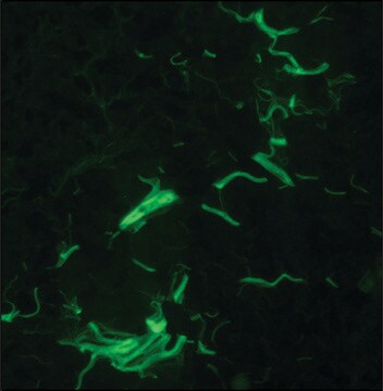
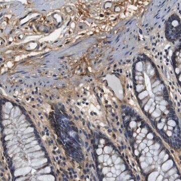
Collagen Type I (cat. # AB755P) staining pattern/morphology in rat skin. Tissue is pre-treated with Citrate pH 6.0, antigen retrieval. Immunoreactivity is seen fiber-staining on paraffin section.

Rat Skin

Forme physique

L'anticorps purifié est fourni dans un tampon contenant un mélange constitué de citrate 0,1 M, de phosphate de potassium 0,1 M, à un pH de 7,2 à 7,4, et de 10 µg/ml d'antibiotiques et d'antimycotiques

Remarque sur l'analyse


Autres remarques

Concentration : pour connaître la concentration spécifique du lot, voir le certificat d'analyse.

Informations légales

CHEMICON is a registered trademark of Merck KGaA, Darmstadt, Germany

Not finding the right product?  

Try our Outil de sélection de produits.

Code de la classe de stockage

13 - Non Combustible Solids

Classe de danger pour l'eau (WGK)

WGK 1

Point d'éclair (°F)

Not applicable

Point d'éclair (°C)

Not applicable


Certificats d'analyse (COA)

Recherchez un Certificats d'analyse (COA) en saisissant le numéro de lot du produit. Les numéros de lot figurent sur l'étiquette du produit après les mots "Lot" ou "Batch".

Déjà en possession de ce produit ?

Retrouvez la documentation relative aux produits que vous avez récemment achetés dans la Bibliothèque de documents.

Consulter la Bibliothèque de documents

CCN3/CCN2 regulation and the fibrosis of diabetic renal disease.
Riser BL, Najmabadi F, Perbal B, Rambow JA, Riser ML, Sukowski E, Yeger H, Riser SC, Peterson DR
Journal of Cell Communication and Signaling null
Thy-1 expression by cardiac fibroblasts: lack of association with myofibroblast contractile markers.
Francois Hudon-David,Fatiha Bouzeghrane,Patrick Couture,Gaetan Thibault
Journal of Molecular and Cellular Cardiology null
Intraglomerular pressure and mesangial stretching stimulate extracellular matrix formation in the rat.
Riser, B L, et al.
The Journal of Clinical Investigation, 90, 1932-1943 (1992)
Partial hepatectomy-induced regeneration accelerates reversion of liver fibrosis involving participation of hepatic stellate cells.
Suarez-Cuenca, Juan A, et al.
Exp. Biol. Med, 233, 827-839 (2008)
Alan S Liu et al.
Scientific reports, 6, 33919-33919 (2016-09-28)
The biomechanical behavior of tissues under mechanical stimulation is critically important to physiological function. We report a combined experimental and modeling study of bioengineered 3D smooth muscle microtissues that reveals a previously unappreciated interaction between active cell mechanics and the

Notre équipe de scientifiques dispose d'une expérience dans tous les secteurs de la recherche, notamment en sciences de la vie, science des matériaux, synthèse chimique, chromatographie, analyse et dans de nombreux autres domaines..

Contacter notre Service technique