Accéder au contenu
MilliporeSigma
Toutes les photos(1)

Key Documents

345860

Sigma-Aldrich

Anti-Glial Fibrillary Acidic Protein Rat mAb (2.2B10)

liquid, clone 2.2B10, Calbiochem®

Synonyme(s) :

Anti-GFAP

Se connecterpour consulter vos tarifs contractuels et ceux de votre entreprise/organisme


About This Item

Code UNSPSC :
12352203
Nomenclature NACRES :
NA.41

Source biologique

rat

Niveau de qualité

Forme d'anticorps

purified antibody

Type de produit anticorps

primary antibodies

Clone

2.2B10, monoclonal

Forme

liquid

Contient

≤0.1% sodium azide as preservative

Espèces réactives

bovine, rat, mouse, human

Fabricant/nom de marque

Calbiochem®

Conditions de stockage

OK to freeze
avoid repeated freeze/thaw cycles

Isotype

IgG2a

Conditions d'expédition

wet ice

Température de stockage

−20°C

Modification post-traductionnelle de la cible

unmodified

Informations sur le gène

human ... GCM1(8521)

Description générale

Purified rat monoclonal antibody. Recognizes the ~55 kDa glial fibrillary acidic protein (GFAP).
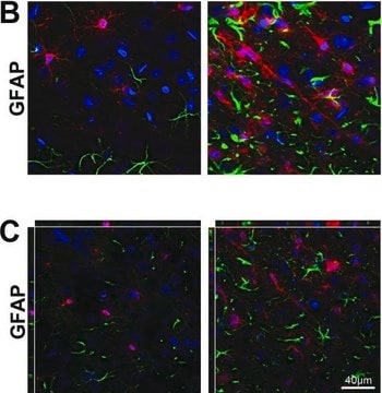
Recognizes GFAP in astrocytes induced by a variety of CNS injuries in human and rat brain tissues.
This Anti-Glial Fibrillary Acidic Protein Rat mAb (2.2B10) is validated for use in ELISA, Immunoblotting, Frozen Sections, IP, Paraffin Sections for the detection of Glial Fibrillary Acidic Protein.

Immunogène

Bovine Brain
bovine GFAP

Application

ELISA (0.1-0.5 µg/ml)

Immunoblotting (0.1-0.5 µg/ml)

Frozen Sections (10-50 µg/ml)

Immunoprecipitation (2-5 µg/ml)

Paraffin Sections (10-50 µg/ml, see comments)

Conditionnement

Please refer to vial label for lot-specific concentration.

Avertissement

Toxicity: Standard Handling (A)

Forme physique

In PBS.

Reconstitution

Following initial thaw, aliquot and freeze (-20°C).

Remarque sur l'analyse

Positive Control
Human or rat brain tissues

Autres remarques

Product is not to be used for animal treatment, in vivo research, or in any other contact procedure with livestock. Stains reactive rodent and human brain astrocytes induced by a variety of central nervous system injuries. This antibody is suitable for immunohistochemical staining of Bouin′s-fixed, frozen, or paraffin-embedded tissue sections. Variables associated with assay conditions will dictate the proper working dilution.
Tohyama, T., et al. 1993. Am. J. Pathol.142, 871.
Lee, V.M., et al. 1984. J. Neurochem. 42, 25.

Informations légales

CALBIOCHEM is a registered trademark of Merck KGaA, Darmstadt, Germany

Vous ne trouvez pas le bon produit ?  

Essayez notre Outil de sélection de produits.

Code de la classe de stockage

11 - Combustible Solids

Classe de danger pour l'eau (WGK)

WGK 1

Point d'éclair (°F)

Not applicable

Point d'éclair (°C)

Not applicable


Certificats d'analyse (COA)

Recherchez un Certificats d'analyse (COA) en saisissant le numéro de lot du produit. Les numéros de lot figurent sur l'étiquette du produit après les mots "Lot" ou "Batch".

Déjà en possession de ce produit ?

Retrouvez la documentation relative aux produits que vous avez récemment achetés dans la Bibliothèque de documents.

Consulter la Bibliothèque de documents

Flora Guillot et al.
Journal of neuroinflammation, 12, 130-130 (2015-07-05)
Astrocytes, the most abundant cell population in mammal central nervous system (CNS), contribute to a variety of functions including homeostasis, metabolism, synapse formation, and myelin maintenance. White matter (WM) reactive astrocytes are important players in amplifying autoimmune demyelination and may
Michael S Petrik et al.
Neuromolecular medicine, 9(1), 83-100 (2006-11-23)
Gulf War illness (GWI) affects a significant percentage of veterans of the 1991 conflict, but its origin remains unknown. Associated with some cases of GWI are increased incidences of amyotrophic lateral sclerosis and other neurological disorders. Whereas many environmental factors
Marco Foddis et al.
Journal of cerebral blood flow and metabolism : official journal of the International Society of Cerebral Blood Flow and Metabolism, 40(2), 276-287 (2019-09-25)
Brain collateral circulation is an essential compensatory mechanism in response to acute brain ischemia. To study the temporal evolution of brain macro and microcollateral recruitment and their reciprocal interactions in response to different ischemic conditions, we applied a combination of
Tadasuke Tominaga et al.
PloS one, 14(3), e0213673-e0213673 (2019-03-12)
Primary and secondary traumatic brain injury (TBI) can cause tissue damage by inducing cell death pathways including apoptosis, necroptosis, and autophagy. However, similar pathways can also lead to senescence. Senescent cells secrete senescence-associated secretory phenotype proteins following persistent DNA damage
Jacob B Ruden et al.
Frontiers in molecular neuroscience, 14, 777049-777049 (2021-12-14)
N-methyl-D-aspartate (NMDA) receptors are critical for higher-order nervous system function, but in previously published protocols to convert human induced pluripotent stem cells (iPSCs) to mature neurons, functional NMDA receptors (NMDARs) are often either not reported or take an extended time

Notre équipe de scientifiques dispose d'une expérience dans tous les secteurs de la recherche, notamment en sciences de la vie, science des matériaux, synthèse chimique, chromatographie, analyse et dans de nombreux autres domaines..

Contacter notre Service technique