1.00485
Masson-Goldner staining kit
for the visualization of connective tissue with trichromic staining
Se connecterpour consulter vos tarifs contractuels et ceux de votre entreprise/organisme
About This Item
Code UNSPSC :
41116124
Nomenclature NACRES :
NA.41
Produits recommandés
Niveau de qualité
IVD
for in vitro diagnostic use
Application(s)
clinical testing
diagnostic assay manufacturing
hematology
histology
Température de stockage
15-25°C
Catégories apparentées
Description générale
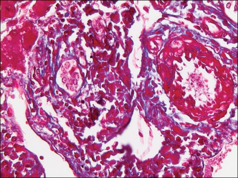
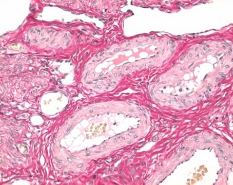
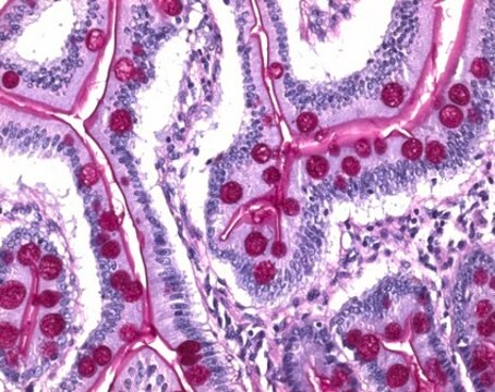
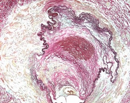
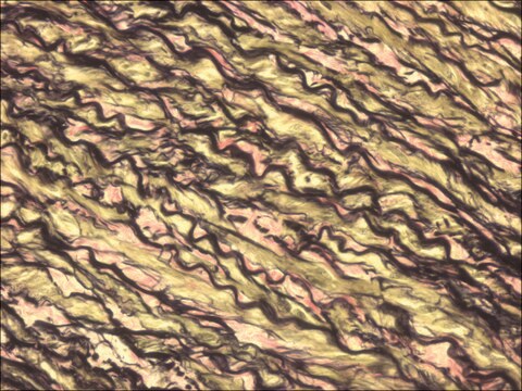
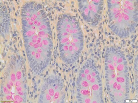
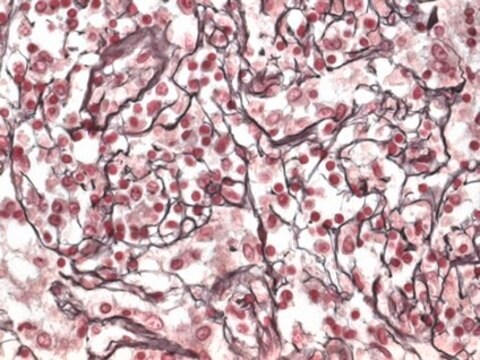
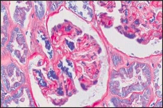
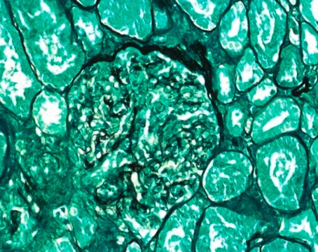
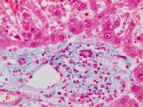
The Masson-Goldner staining kit - for the visualization of connective tissue with trichromic staining, is used for human-medical cell diagnosis and serves the purpose of the histological investigation of sample material of human origin. Using a combination of three different staining solutions, muscle fibers, collagenous fibers, fibrin and erythrocytes can be selectively visualized.
The original methods were primarily used to differentiate collagenous and muscle fibers. The stains used have different molecular sizes and enable the individual tissues to be stained differentially.
The Masson-Goldner staining technique can be carried out using formalin fixed material. Subsequent to staining the nucleus with Weigert′s iron hematoxylin, components such as muscle, cytoplasm and erythrocytes are stained with azophloxin and orange G solution. Connective tissue is then counter stained using light green SF solution.
The package is sufficient for 400 - 500 applications. The product is registered as IVD and CE product and can be used in diagnostics and laboratory accreditation. For more details, please see instructions for use (IFU). The IFU can be downloaded from this webpage.
Remarque sur l'analyse
Suitability for microscopy (Tissue section): passes test
Nuclei: dark brown to black
Cytoplasm: brick red
musculature (muscles): bright red
Connective tissue: green
acid mucosubstances: green
Erythrocytes: bright orange
Nuclei: dark brown to black
Cytoplasm: brick red
musculature (muscles): bright red
Connective tissue: green
acid mucosubstances: green
Erythrocytes: bright orange
Mention d'avertissement
Danger
Mentions de danger
Conseils de prudence
Classification des risques
Eye Dam. 1 - Skin Irrit. 2
Code de la classe de stockage
8A - Combustible, corrosive hazardous materials
Classe de danger pour l'eau (WGK)
WGK 2
Certificats d'analyse (COA)
Recherchez un Certificats d'analyse (COA) en saisissant le numéro de lot du produit. Les numéros de lot figurent sur l'étiquette du produit après les mots "Lot" ou "Batch".
Déjà en possession de ce produit ?
Retrouvez la documentation relative aux produits que vous avez récemment achetés dans la Bibliothèque de documents.
Les clients ont également consulté
Aurora Garre et al.
Clinical, cosmetic and investigational dermatology, 11, 253-263 (2018-06-09)
With age, decreasing dermal levels of proteoglycans, collagen, and elastin lead to the appearance of aged skin. Oxidation, largely driven by environmental factors, plays a central role. The aim of this study was to assess the antiaging efficacy of a
B Shen et al.
Nan fang yi ke da xue xue bao = Journal of Southern Medical University, 41(7), 1107-1113 (2021-07-27)
To investigate the effect of ginsenoside Rh2 (G-Rh2) on renal fibrosis and cell apoptosis in rats with diabetic nephropathy (DN) and explore its possible mechanism. Thirty male SD rats were randomized equally into control group, DN group and ginsenoside Rh2
Álvaro Santana-Garrido et al.
Journal of physiology and biochemistry (2022-08-10)
Arterial hypertension (AH) leads to oxidative and inflammatory imbalance that contribute to fibrosis development in many target organs. Here, we aimed to highlight the harmful effects of severe AH in the cornea. Our experimental model was established by administration of
Notre équipe de scientifiques dispose d'une expérience dans tous les secteurs de la recherche, notamment en sciences de la vie, science des matériaux, synthèse chimique, chromatographie, analyse et dans de nombreux autres domaines..
Contacter notre Service technique