Accéder au contenu
MilliporeSigma
Toutes les photos(1)

Key Documents

04-1530

Sigma-Aldrich

Anti-MDM2 Antibody, clone 3G9

clone 3G9, from mouse

Synonyme(s) :

Double minute 2 protein, Mdm2 p53 binding protein homolog (mouse), Mdm2, transformed 3T3 cell double minute 2, p53 binding protein, Mdm2, transformed 3T3 cell double minute 2, p53 binding protein (mouse), Oncoprotein Mdm2, double minute 2, human homolog

Se connecterpour consulter vos tarifs contractuels et ceux de votre entreprise/organisme


About This Item

Code UNSPSC :
12352203
eCl@ss :
32160702
Nomenclature NACRES :
NA.41

Source biologique

mouse

Niveau de qualité

Forme d'anticorps

purified antibody

Type de produit anticorps

primary antibodies

Clone

3G9, monoclonal

Espèces réactives

human

Réactivité de l'espèce (prédite par homologie)

mouse (based on 100% sequence homology)

Conditionnement

antibody small pack of 25 μL

Technique(s)

immunocytochemistry: suitable
immunohistochemistry: suitable
immunoprecipitation (IP): suitable
western blot: suitable

Numéro d'accès NCBI

Numéro d'accès UniProt

Conditions d'expédition

ambient

Modification post-traductionnelle de la cible

unmodified

Informations sur le gène

human ... MDM2(4193)
mouse ... Mdm2(17246)

Description générale

MDM2 is over-expressed in many tumors. Its principal function is the ubiquitination and degradation of p53 tumor suppressor protein. This monoclonal antibody also recognizes a peptide epitope around Thr-216 of murine MDM2 when Thr-216 is unphosphorylated. SMP14 also cross reacts with some cytokeratins (6, 14 & 16). This is only a problem when working with certain epithelial cells and not fibroblasts.

Spécificité

This antibody recognizes MDM2.

Immunogène

Epitope: Unknown
His-tagged recombinant protein corresponding to human MDM2.

Application

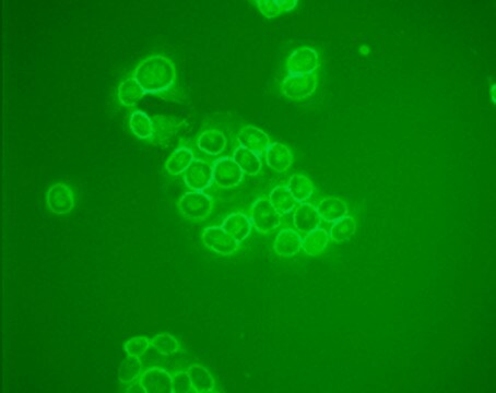
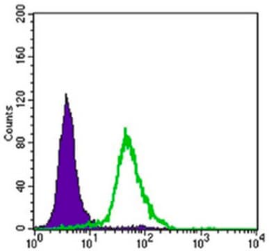
Anti-MDM2 Antibody, clone 3G9 is a Mouse Monoclonal Antibody for detection of MDM2 also known as Double minute 2 protein or Mdm2 p53 binding protein homolog & has been validated in WB, IP, ICC & IHC.
Immunoprecipitation Analysis: A representative lot was used by an independent laboratory for this application. (Changgong, L., et al. (2002). Molecular and Cellular Biology. 22(21):7562-7571.)
Research Category
Epigenetics & Nuclear Function
Research Sub Category
RNA Metabolism & Binding Proteins

Ubiquitin & Ubiquitin Metabolism

Qualité

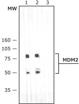
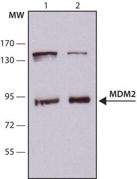
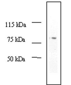
Evaluated by Western Blot in MCF-7 cell lysate.

Western Blot Analysis: 0.5 µg/ml of this antibody detected MDM2 on 10 µg of MCF-7 cell lysate.

Description de la cible

~ 75 kDa

Forme physique

Format: Purified
Protein G Purified
Purified mouse monoclonal IgGκ in buffer containing 0.1 M Tris-Glycine (pH 7.4, 150 mM NaCl) with 0.05% sodium azide.

Stockage et stabilité

Stable for 1 year at 2-8°C from date of receipt.

Remarque sur l'analyse

Control
MCF-7 cell lysate

Autres remarques

Concentration: Please refer to the Certificate of Analysis for the lot-specific concentration.

Clause de non-responsabilité

Unless otherwise stated in our catalog or other company documentation accompanying the product(s), our products are intended for research use only and are not to be used for any other purpose, which includes but is not limited to, unauthorized commercial uses, in vitro diagnostic uses, ex vivo or in vivo therapeutic uses or any type of consumption or application to humans or animals.

Vous ne trouvez pas le bon produit ?  

Essayez notre Outil de sélection de produits.

Code de la classe de stockage

12 - Non Combustible Liquids

Classe de danger pour l'eau (WGK)

WGK 1

Point d'éclair (°F)

Not applicable

Point d'éclair (°C)

Not applicable


Certificats d'analyse (COA)

Recherchez un Certificats d'analyse (COA) en saisissant le numéro de lot du produit. Les numéros de lot figurent sur l'étiquette du produit après les mots "Lot" ou "Batch".

Déjà en possession de ce produit ?

Retrouvez la documentation relative aux produits que vous avez récemment achetés dans la Bibliothèque de documents.

Consulter la Bibliothèque de documents

Zhenzhen Liu et al.
Chemical biology & drug design, 85(4), 411-417 (2014-09-18)
In this article, we describe a no-wash small-molecule fluorescent probe for detecting and imaging p53-MDM2 protein-protein interaction based on an environment-sensitive fluorescent turn-on mechanism. After extensive biological examination, this probe L1 exhibited practical activity and selectivity in vitro and in
Lenka Kyjacova et al.
Oncogene, 40(47), 6494-6512 (2021-10-07)
Expression of the immediate-early response gene IER2 has been associated with the progression of several types of cancer, but its functional role is poorly understood. We found that increased IER2 expression in human melanoma is associated with shorter overall survival
R C Heijkants et al.
Oncogenesis, 7(3), 33-33 (2018-03-30)
Uveal melanoma (UM) is the most frequent ocular cancer in adults, accounting for ~5% of the total melanoma incidence. Although the primary tumor is well treatable, patients frequently develop metastases for which no curative therapy exists. Highly activated protein kinase
Xiaomu Li et al.
Nature communications, 7, 11740-11740 (2016-06-07)
Mitochondrial metabolism is pivotal for glucose-stimulated insulin secretion (GSIS) in pancreatic β-cells. However, little is known about the molecular machinery that controls the homeostasis of intermediary metabolites in mitochondria. Here we show that the activation of p53 in β-cells, by
Chloé Prunier et al.
NPJ breast cancer, 7(1), 140-140 (2021-10-29)
Reactivation of dormant cancer cells can lead to cancer relapse, metastasis, and patient death. Dormancy is a nonproliferative state and is linked to late relapse and death. No targeted therapy is currently available to eliminate dormant cells, highlighting the need

Notre équipe de scientifiques dispose d'une expérience dans tous les secteurs de la recherche, notamment en sciences de la vie, science des matériaux, synthèse chimique, chromatographie, analyse et dans de nombreux autres domaines..

Contacter notre Service technique