Skip to Content
MilliporeSigma
All Photos(3)

Key Documents

SAB4300525

Sigma-Aldrich

Anti-GAP43 (Ab-41) antibody produced in rabbit

affinity isolated antibody

Synonym(s):

Anti-B-50 antibody produced in rabbit, Anti-PP46 antibody produced in rabbit, Anti-growth associated protein 43 antibody produced in rabbit

Sign Into View Organizational & Contract Pricing


About This Item

MDL number:
UNSPSC Code:
12352203
NACRES:
NA.41

biological source

rabbit

conjugate

unconjugated

antibody form

affinity isolated antibody

antibody product type

primary antibodies

clone

polyclonal

form

buffered aqueous solution

mol wt

~43 kDa

species reactivity

rat, human, mouse

concentration

1 mg/mL

technique(s)

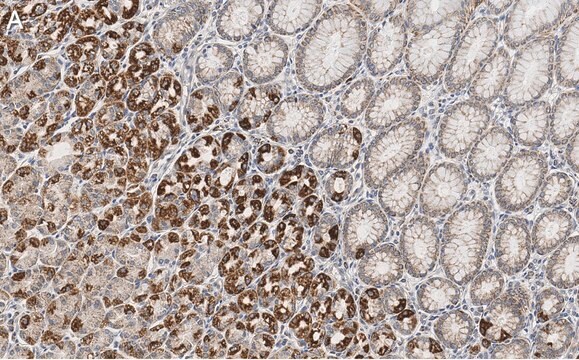
immunohistochemistry (formalin-fixed, paraffin-embedded sections): 1:50-1:100
indirect immunofluorescence: 1:100-1:200
western blot: 1:500-1:1000

isotype

IgG

immunogen sequence

(Q-A-S-F-R)

NCBI accession no.

UniProt accession no.

shipped in

wet ice

storage temp.

−20°C

target post-translational modification

unmodified

Gene Information

human ... GAP43(2596)

General description

Growth associated protein-43 (GAP-43) is a phosphoprotein associated with membrane. It is located on chromosome 3q13. During neural development and axonal regeneration, GAP43 is upregulated in neuronal growth cones and Schwann cell precursors.

Immunogen

Peptide sequence around aa. 38-43 (Q-A-S-F-R), according to the protein GAP43.

Biochem/physiol Actions

Growth associated protein-43 (GAP-43) participates in neurite outgrowth, growth cone navigation, neurotransmission, stabilization of axonal branches and synaptic plasticity. In humans and mice, GAP43 is recognized as a possible gene for autism and autistic-like disorders.

Features and Benefits

Evaluate our antibodies with complete peace of mind. If the antibody does not perform in your application, we will issue a full credit or replacement antibody. Learn more.

Target description

GAP43 encoded by this gene has been termed a 'growth' or 'plasticity' protein because it is expressed at high levels in neuronal growth cones during development and axonal regeneration. This protein is considered a crucial component of an effective regenerative response in the nervous system. Alternatively spliced transcript variants encoding distinct isoforms have been found for this gene.

Physical form

Solution in phosphate-buffered saline containing 0.02% sodium azide and 50% glycerol

Disclaimer

Unless otherwise stated in our catalog or other company documentation accompanying the product(s), our products are intended for research use only and are not to be used for any other purpose, which includes but is not limited to, unauthorized commercial uses, in vitro diagnostic uses, ex vivo or in vivo therapeutic uses or any type of consumption or application to humans or animals.

Not finding the right product?  

Try our Product Selector Tool.

Storage Class Code

10 - Combustible liquids

WGK

WGK 1

Flash Point(F)

Not applicable

Flash Point(C)

Not applicable


Certificates of Analysis (COA)

Search for Certificates of Analysis (COA) by entering the products Lot/Batch Number. Lot and Batch Numbers can be found on a product’s label following the words ‘Lot’ or ‘Batch’.

Already Own This Product?

Find documentation for the products that you have recently purchased in the Document Library.

Visit the Document Library

A rare 3q13.31 microdeletion including GAP43 and LSAMP genes
Gimelli S, et al.
Molecular Cytogenetics, 6(1), 52-52 (2013)
Víctor Carriel et al.
Journal of tissue engineering and regenerative medicine, 11(2), 553-563 (2014-08-02)
Nerve conduits are promising alternatives for repairing nerve gaps; they provide a close microenvironment that supports nerve regeneration. In this sense, histological analysis of axonal growth is a determinant to achieve successful nerve regeneration. To evaluate this process, the most-used
Wei-Shen Chen et al.
Modern pathology : an official journal of the United States and Canadian Academy of Pathology, Inc, 27(2), 184-193 (2013-07-28)
The malignant peripheral nerve sheath tumor is a relatively uncommon type of soft tissue sarcoma arising from a peripheral nerve or extraneural soft tissues and showing nerve sheath differentiation. The diagnosis of malignant peripheral nerve sheath tumor is one of
Ryan L O'Hare Doig et al.
Frontiers in molecular neuroscience, 13, 85-85 (2020-07-17)
Reducing the extent of secondary degeneration following spinal cord injury (SCI) is necessary to preserve function, but treatment options have thus far been limited. A combination of the ion channel inhibitors Lomerizine (Lom), YM872 and oxATP, to inhibit voltage-gated Ca2+
Nuttapong Yawoot et al.
Journal of cellular physiology, 237(3), 1818-1832 (2021-11-27)
Even though astrocytes have been widely reported to support several brain functions, studies have emerged that they exert deleterious effects on the brain after ischemia and reperfusion (I/R) injury. The present study investigated the neuroprotective effects of melatonin on the

Our team of scientists has experience in all areas of research including Life Science, Material Science, Chemical Synthesis, Chromatography, Analytical and many others.

Contact Technical Service